术中超声诊断在肝脏肿瘤手术中的作用
作者:于健春 钟守先 朱预
单位:中国医学科学院 中国协和医科大学 协和医院外科,北京 100730
关键词:术中超声;肝脏肿瘤;手术
术中超声诊断在肝脏肿瘤手术中的作用 摘要 目的 研究术中超声对肝脏肿瘤患者手术治疗的作用。方法 应用5.0-MHz特殊超声探头,对49例患肝脏肿瘤或其他疾病可疑肝脏转移肿瘤的患者在开腹探查手术中进行术中超声检查。根据术前影像学检查(包括体外经皮超声、核磁共振成像和计算机断层扫描)及术中评定(包括肉眼观察、双手触诊及术中超声),对术前影像学检查与术中超声方法进行对比分析。 结果 术中超声、体外经皮超声、计算机断层扫描、核磁共振成像的敏感性分别为100%(23/23)、74%(17/23)、74%(14/19)和75%(6/8),特异性分别为100%(26/26)、100%(26/26)、93%(14/15)和67%(2/3)。7例(14%)肝脏肿瘤患者经术中望诊、触诊均未发现病灶,仅靠术中超声检查才得以确定。结论 术中超声是检查发现肝脏肿瘤的最敏感、特异的方法,它有助于肝脏肿瘤,尤其是肝脏隐匿性或微小病灶(<2 cm)的手术治疗。
, http://www.100md.com
中图号 R445.1 R657.3
Evaluation of Intraoperative Ultrasonography for Hepatic Neoplasm in Surgery
Yu Jianchun Zhong Shouxian Zhu Yu
(Department of General Surgery,PUMC Hospital,CAMS and PUMC,Beijing 100730)
Objective The purpose of this study was to evaluate the impact of intraoperative ultrasound (IOUS) on the management of patients with neoplasms of the liver.Methods Forty-nine patients received operations for liver tumors were examined intraoperatively with 5.0 MHz special ultrasound transducers during surgical exploration of the abdomen.Preoperative imaging studies including percutaneous ultrasound (n=49),magnetic resonance imaging (n=11),and computed tomography (n=34) were taken to compare with intraoperative ultrasonography for the evaluation.Results Sensitivity for detection of hepatic neoplasms showed in intraoperative ultrasound ,percutaneous ultrasound,magnetic resonance imaging and computed tomography as 100%(23/23),74%(17/23),74%(14/19),and 75%(6/8) respectively.Specificity showed 100%(26/26),100%(26/26),93%(14/15),and 67(2/3) respectively.Conclusions Intraoperative ultrasound was the most sensitive and specific method for detection and surgery of liver neoplasms,especially the occult neoplasms and small size lesion(<2 cm).
, 百拇医药
Key words intraoperative ultrasound;hepatic neoplsm;surgery
现代术中超声(intraoperative ultrasound,IOUS)在80年代初首先被证明可用于胆道和胰腺手术[1,2]。此后,Makuuchi等[3,4]报道在肝脏手术中应用术中超声可清晰显示肝脏肿瘤图象,从而安全、有效地进行手术切除。尽管无创性的影像学技术,如体外经皮超声、计算机断层扫描(CT)及核磁共振成像(MRI)已广泛用于肝脏肿瘤的诊断,但其阳性发现率不超过60%~80%[5~8]。精密的超声附件使术中超声更为有效和方便,在许多普通外科疾病,尤其是在肝脏肿瘤治疗中,术中超声已为普外科医师所认识并采用[9~12]。本研究根据前瞻性的诊断和回顾性资料分析,探讨术中超声在肝脏手术中的作用。
1 材料和方法
, http://www.100md.com
本科住院患者49例,入选标准为已知有肝脏病灶或可疑有肝脏病灶,后者如肝转移癌可能性大的胃癌或结肠癌患者。
术前准备包括病史采集、体格检查、生化指标、胸部X线拍片、癌胚抗原等常规检查。术前影像学检查包括体外经皮超声(n=49),核磁共振成像(n=11)及计算机断层扫描(n=34)。
术中评定通过开腹探查时的肉眼观察、双手触诊及术中超声检查确定。各种术前影像学检查均按公式进行敏感性和特异性评定,并将术前影像学检查与术中评定进行对比分析。
应用特殊的术中超声仪(Sonoline SI-250,德国西门子公司),由一名有经验的外科医师持5.0-MHz线阵超声探头(图1)进行术中超声检查。I型或T型两种术中超声探头极为小巧,可放在触诊探查两指间。因术中超声探头及其导线用气体消毒需48 h,故本研究购用一次性无菌塑料套套住探头,以便及时用于手术患者。超声探头晶体面与无菌塑料套之间用超声耦合剂介导。
, 百拇医药
图 1 5.0-MHz线阵术中超声探头
Fig 1 A 5.0-MHz linear array
transducer
整个肝脏以纵向和横向扫描。探头由右肝外侧开始,逐步移向左侧圆韧带,同样方法扫描左肝外侧叶。探头与肝脏之间由腹腔体液或无菌生理盐水接触介导。该操作需使手术时间延长10~15 min。无并发症及无菌区污染发生。
所有数据均用STATVIEW标准统计软件在苹果电脑Power Macintosh 7220上进行统计分析。敏感性和特异性均用卡方检验。
2 结果
经手术、术中超声和病理证实,49例患者中23例患肝脏原发性或继发性肿瘤,其中15例为恶性肿瘤(原发性肝癌9例,转移癌6例),8例为良性肿瘤(血管瘤3例,肝囊肿4例,胆管腺瘤1例)。
, http://www.100md.com
术中超声与术前不同影像学检查结果总体比较表明,术中超声最为敏感和特异。术中超声、体外经皮超声、计算机断层扫描、核磁共振成像的敏感性分别为100%(23/23)、74%(17/23)、74%(14/19)和75%(6/8);术中超声与核磁共振成像比较差异有显著意义(P<0.05),与体外经皮超声、计算机断层扫描比较差异有非常显著意义(P<0.01)。上述四种影像学诊断的特异性分别为100%(26/26)、100%(26/26)、93%(14/15)和67%(2/3),术中超声与核磁共振成像比较差异亦有显著性意义(P<0.05)。
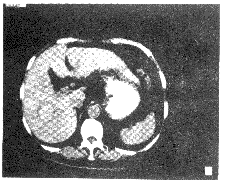
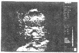
9例患者(18%)术前影像学检查未见可疑病灶,经术中超声发现肿瘤。7例患者(14%)经术中望、触诊均未发现肿瘤病灶,但经术中超声检查发现肝脏肿瘤。其中2例女性患者(59及42岁)术前已经CT、血管造影及MRI诊为右肝细胞肝癌,术中望诊、触诊均未发现病灶,但术中超声清晰显示了肿瘤及其周围血管和胆管结构(图2),从而使肿瘤得以顺利切除。1例男性患者(31岁)仅于术前体外超声发现左肝叶一1.6 cm×1.0 cm病灶,而CT和术中望、触诊均未发现病灶,后经术中超声发现病灶并予切除,病理诊断为胆管腺瘤。其余4例患者均经术中超声发现为0.6~2 cm小病灶。

, http://www.100md.com
图2 59岁女性患者术前CT显示右肝癌,术中超声显示肿瘤及其周围血管和胆管结构
Fig 2 A 59-year-old woman with right hepatic carcinoma imaged by CT (a).The tumor with surrounding vessels and biliary structures was identified only by intraoperative ultrasound during exploration (b)
3 讨论
术前诊断肝脏肿瘤最常用的影像学方法为传统的体外经皮超声、计算机断层扫描及核磁共振成像。尽管各种影像的实际分辨率均有所提高,但其敏感性仍为40%~85%[5~9,12]。应用CT血管成像及使用新型造影剂改良的标准普通CT可使敏感性提高到90%~96%[13,14]。有人报道以上3种影像学方法总的敏感性仅为77%[5],原因在于这3种影像学方法对小于2 cm病灶的探测有限。经皮腹腔超声时,由于皮肤、皮下组织、肌肉、肋骨及肠道气体导致的声波衰减和散射,在很大程度上妨碍了理想的超声成像。而且,体外超声需较大的声波以便穿透距离相对较远的腹腔内脏器,因而需要较低频率的探头,而较低频率的探头又会直接影响图象的分辨率和质量。
, 百拇医药
近年来人们认识到,术中超声可以发现许多其他影像学检查无法发现的病灶[8,11~16]。术中超声这种特殊的高级声像仪器使手术中快速、准确成像成为可能,有经验的外科医师能够很快学会并运用这种有用的工具。术中超声虽与传统超声的诊断标准相似,但它却具有可直接或以最短距离置于脏器表面的优点。
本研究在腹部探查时以5.0MHz的I型或T型探头进行术中超声,证明以往报道的术中超声的优越性是经得起手术考验的。术中超声的影像资料有助于分辨肝脏中正常或异常的胆管或血管的解剖结构,确定已知的肿瘤及其他隐匿的肿瘤,尤其是可疑病灶、微小病灶(<2 cm)或术前影像学检查发现、但术中无法扪及的病灶。
于健春为通讯作者
参考文献
1 Sigel B,Coelho JV,Nyhus LM,et al.Comparison of cholangiography and ultrasonography in the operative screening of the common bile duct.World J Surg,1982,6:440~444
, http://www.100md.com
2 Sigel B,Coelho JC,Nyhus LM,et al.Detection of pancreatic tumors by ultrasound during surgery.Arch Surg,1982,117:1058~1061
3 Makuuchi M,Hasegawa H,Yamazaki S.Intraoperative ultrasonic examination for hepatectomy.Jpn J Clin Oncol,1981,11:367~369
4 Gozzetti G,Mazziotti A,Bolondi L,et al.Intraoperative ultrasonography in surgery for liver tumors.Surgery,1986,99:523~529
5 Wernecke K,Rummeny E,Bongartz G, et al.Detection of hepatic masses in patients with carcinoma:comparative sensitivities of sonography,CT,and MR imaging.AJR,1991,157:731~739
, 百拇医药
6 Clarke MP,Kane RA,Steele G Jr,et al.Prospective comparison of preoperative imaging and intraoperative ultrasonography in the detection of liver tumors.Surgery,1989,106:849~855
7 Cater R,Hemingway D,Cook TG,et al.A prospective study of six methods for detection of hepatic colorectal metastases.Ann R Coll Surg Engl,1996,78(1):27~30
8 Hagspiel KD,Neidl KF,Eichenberger AC,et al.Detection of liver metastases:comparison of superparamagnetic iron oxide-enhanced and unenhanced MR imaging at 1.5 T with dynamic CT,intraoperative US,and percutaneous US.Radiology,1995,196(2):471~478
, http://www.100md.com
9 Soyer P,Levesque M,Elias D,et al.Detection of liver metastases from colorectal cancer:comparison of intraoperative US and CT during arterial photography.Radiology,1991,183:541~544
10 Cerri LM,Cerri GG.Intraoperative ultrasonography of liver,bile ducts and pancreas.Rev Paul Med,1996,114(4):1196~1207
11 Kane RA,Hughes LA,Cua EJ,et al.The impact of intraoperative ultrasonography on surgery for liver neoplasms.J Ultrasound Med,1994,13:1~6
, 百拇医药
12 Machi J,Isomoto H,Kurohiji T,et al.Accuracy of intraoperative ultrasonography in diagnosing liver metastasis from colorectal cancer:evaluation with postoperative follow-up results.World J Surg,1991,15:551~556
13 Staren ED,Gambla M,Deziel DJ,et al.Intraoperative ultrasound in the management of liver neoplasms.Am Surg,1997,63(7):591~597
14 Knol JA,Marn CS,Francis IR,et al.Comparisons of dynamic infusion and delayed computed tomography,intraoperative ultrasound,and palpation in the diagnosis of liver metastases.Am J Surg,1993,165:81~87
, 百拇医药
15 Ravikumar TS,Buenaventura S,Salem RR,et al.Intrasonography of liver:detection of occult liver tumors and treatment by cryosurgery.Cancer Detect Prev,1994,18:131~138
16 Hanna SS,Nam R,Leonhardt C.Liver resection by ultrsonic dissection and intraoperative ultrasonog-raphy.HPB Surg,1996,9(3):121~128
(1999-02-01 收稿), 百拇医药
单位:中国医学科学院 中国协和医科大学 协和医院外科,北京 100730
关键词:术中超声;肝脏肿瘤;手术
术中超声诊断在肝脏肿瘤手术中的作用 摘要 目的 研究术中超声对肝脏肿瘤患者手术治疗的作用。方法 应用5.0-MHz特殊超声探头,对49例患肝脏肿瘤或其他疾病可疑肝脏转移肿瘤的患者在开腹探查手术中进行术中超声检查。根据术前影像学检查(包括体外经皮超声、核磁共振成像和计算机断层扫描)及术中评定(包括肉眼观察、双手触诊及术中超声),对术前影像学检查与术中超声方法进行对比分析。 结果 术中超声、体外经皮超声、计算机断层扫描、核磁共振成像的敏感性分别为100%(23/23)、74%(17/23)、74%(14/19)和75%(6/8),特异性分别为100%(26/26)、100%(26/26)、93%(14/15)和67%(2/3)。7例(14%)肝脏肿瘤患者经术中望诊、触诊均未发现病灶,仅靠术中超声检查才得以确定。结论 术中超声是检查发现肝脏肿瘤的最敏感、特异的方法,它有助于肝脏肿瘤,尤其是肝脏隐匿性或微小病灶(<2 cm)的手术治疗。
, http://www.100md.com
中图号 R445.1 R657.3
Evaluation of Intraoperative Ultrasonography for Hepatic Neoplasm in Surgery
Yu Jianchun Zhong Shouxian Zhu Yu
(Department of General Surgery,PUMC Hospital,CAMS and PUMC,Beijing 100730)
Objective The purpose of this study was to evaluate the impact of intraoperative ultrasound (IOUS) on the management of patients with neoplasms of the liver.Methods Forty-nine patients received operations for liver tumors were examined intraoperatively with 5.0 MHz special ultrasound transducers during surgical exploration of the abdomen.Preoperative imaging studies including percutaneous ultrasound (n=49),magnetic resonance imaging (n=11),and computed tomography (n=34) were taken to compare with intraoperative ultrasonography for the evaluation.Results Sensitivity for detection of hepatic neoplasms showed in intraoperative ultrasound ,percutaneous ultrasound,magnetic resonance imaging and computed tomography as 100%(23/23),74%(17/23),74%(14/19),and 75%(6/8) respectively.Specificity showed 100%(26/26),100%(26/26),93%(14/15),and 67(2/3) respectively.Conclusions Intraoperative ultrasound was the most sensitive and specific method for detection and surgery of liver neoplasms,especially the occult neoplasms and small size lesion(<2 cm).
, 百拇医药
Key words intraoperative ultrasound;hepatic neoplsm;surgery
现代术中超声(intraoperative ultrasound,IOUS)在80年代初首先被证明可用于胆道和胰腺手术[1,2]。此后,Makuuchi等[3,4]报道在肝脏手术中应用术中超声可清晰显示肝脏肿瘤图象,从而安全、有效地进行手术切除。尽管无创性的影像学技术,如体外经皮超声、计算机断层扫描(CT)及核磁共振成像(MRI)已广泛用于肝脏肿瘤的诊断,但其阳性发现率不超过60%~80%[5~8]。精密的超声附件使术中超声更为有效和方便,在许多普通外科疾病,尤其是在肝脏肿瘤治疗中,术中超声已为普外科医师所认识并采用[9~12]。本研究根据前瞻性的诊断和回顾性资料分析,探讨术中超声在肝脏手术中的作用。
1 材料和方法
, http://www.100md.com
本科住院患者49例,入选标准为已知有肝脏病灶或可疑有肝脏病灶,后者如肝转移癌可能性大的胃癌或结肠癌患者。
术前准备包括病史采集、体格检查、生化指标、胸部X线拍片、癌胚抗原等常规检查。术前影像学检查包括体外经皮超声(n=49),核磁共振成像(n=11)及计算机断层扫描(n=34)。
术中评定通过开腹探查时的肉眼观察、双手触诊及术中超声检查确定。各种术前影像学检查均按公式进行敏感性和特异性评定,并将术前影像学检查与术中评定进行对比分析。

应用特殊的术中超声仪(Sonoline SI-250,德国西门子公司),由一名有经验的外科医师持5.0-MHz线阵超声探头(图1)进行术中超声检查。I型或T型两种术中超声探头极为小巧,可放在触诊探查两指间。因术中超声探头及其导线用气体消毒需48 h,故本研究购用一次性无菌塑料套套住探头,以便及时用于手术患者。超声探头晶体面与无菌塑料套之间用超声耦合剂介导。

, 百拇医药
图 1 5.0-MHz线阵术中超声探头
Fig 1 A 5.0-MHz linear array
transducer
整个肝脏以纵向和横向扫描。探头由右肝外侧开始,逐步移向左侧圆韧带,同样方法扫描左肝外侧叶。探头与肝脏之间由腹腔体液或无菌生理盐水接触介导。该操作需使手术时间延长10~15 min。无并发症及无菌区污染发生。
所有数据均用STATVIEW标准统计软件在苹果电脑Power Macintosh 7220上进行统计分析。敏感性和特异性均用卡方检验。
2 结果
经手术、术中超声和病理证实,49例患者中23例患肝脏原发性或继发性肿瘤,其中15例为恶性肿瘤(原发性肝癌9例,转移癌6例),8例为良性肿瘤(血管瘤3例,肝囊肿4例,胆管腺瘤1例)。
, http://www.100md.com
术中超声与术前不同影像学检查结果总体比较表明,术中超声最为敏感和特异。术中超声、体外经皮超声、计算机断层扫描、核磁共振成像的敏感性分别为100%(23/23)、74%(17/23)、74%(14/19)和75%(6/8);术中超声与核磁共振成像比较差异有显著意义(P<0.05),与体外经皮超声、计算机断层扫描比较差异有非常显著意义(P<0.01)。上述四种影像学诊断的特异性分别为100%(26/26)、100%(26/26)、93%(14/15)和67%(2/3),术中超声与核磁共振成像比较差异亦有显著性意义(P<0.05)。
9例患者(18%)术前影像学检查未见可疑病灶,经术中超声发现肿瘤。7例患者(14%)经术中望、触诊均未发现肿瘤病灶,但经术中超声检查发现肝脏肿瘤。其中2例女性患者(59及42岁)术前已经CT、血管造影及MRI诊为右肝细胞肝癌,术中望诊、触诊均未发现病灶,但术中超声清晰显示了肿瘤及其周围血管和胆管结构(图2),从而使肿瘤得以顺利切除。1例男性患者(31岁)仅于术前体外超声发现左肝叶一1.6 cm×1.0 cm病灶,而CT和术中望、触诊均未发现病灶,后经术中超声发现病灶并予切除,病理诊断为胆管腺瘤。其余4例患者均经术中超声发现为0.6~2 cm小病灶。


, http://www.100md.com
图2 59岁女性患者术前CT显示右肝癌,术中超声显示肿瘤及其周围血管和胆管结构
Fig 2 A 59-year-old woman with right hepatic carcinoma imaged by CT (a).The tumor with surrounding vessels and biliary structures was identified only by intraoperative ultrasound during exploration (b)
3 讨论
术前诊断肝脏肿瘤最常用的影像学方法为传统的体外经皮超声、计算机断层扫描及核磁共振成像。尽管各种影像的实际分辨率均有所提高,但其敏感性仍为40%~85%[5~9,12]。应用CT血管成像及使用新型造影剂改良的标准普通CT可使敏感性提高到90%~96%[13,14]。有人报道以上3种影像学方法总的敏感性仅为77%[5],原因在于这3种影像学方法对小于2 cm病灶的探测有限。经皮腹腔超声时,由于皮肤、皮下组织、肌肉、肋骨及肠道气体导致的声波衰减和散射,在很大程度上妨碍了理想的超声成像。而且,体外超声需较大的声波以便穿透距离相对较远的腹腔内脏器,因而需要较低频率的探头,而较低频率的探头又会直接影响图象的分辨率和质量。
, 百拇医药
近年来人们认识到,术中超声可以发现许多其他影像学检查无法发现的病灶[8,11~16]。术中超声这种特殊的高级声像仪器使手术中快速、准确成像成为可能,有经验的外科医师能够很快学会并运用这种有用的工具。术中超声虽与传统超声的诊断标准相似,但它却具有可直接或以最短距离置于脏器表面的优点。
本研究在腹部探查时以5.0MHz的I型或T型探头进行术中超声,证明以往报道的术中超声的优越性是经得起手术考验的。术中超声的影像资料有助于分辨肝脏中正常或异常的胆管或血管的解剖结构,确定已知的肿瘤及其他隐匿的肿瘤,尤其是可疑病灶、微小病灶(<2 cm)或术前影像学检查发现、但术中无法扪及的病灶。
于健春为通讯作者
参考文献
1 Sigel B,Coelho JV,Nyhus LM,et al.Comparison of cholangiography and ultrasonography in the operative screening of the common bile duct.World J Surg,1982,6:440~444
, http://www.100md.com
2 Sigel B,Coelho JC,Nyhus LM,et al.Detection of pancreatic tumors by ultrasound during surgery.Arch Surg,1982,117:1058~1061
3 Makuuchi M,Hasegawa H,Yamazaki S.Intraoperative ultrasonic examination for hepatectomy.Jpn J Clin Oncol,1981,11:367~369
4 Gozzetti G,Mazziotti A,Bolondi L,et al.Intraoperative ultrasonography in surgery for liver tumors.Surgery,1986,99:523~529
5 Wernecke K,Rummeny E,Bongartz G, et al.Detection of hepatic masses in patients with carcinoma:comparative sensitivities of sonography,CT,and MR imaging.AJR,1991,157:731~739
, 百拇医药
6 Clarke MP,Kane RA,Steele G Jr,et al.Prospective comparison of preoperative imaging and intraoperative ultrasonography in the detection of liver tumors.Surgery,1989,106:849~855
7 Cater R,Hemingway D,Cook TG,et al.A prospective study of six methods for detection of hepatic colorectal metastases.Ann R Coll Surg Engl,1996,78(1):27~30
8 Hagspiel KD,Neidl KF,Eichenberger AC,et al.Detection of liver metastases:comparison of superparamagnetic iron oxide-enhanced and unenhanced MR imaging at 1.5 T with dynamic CT,intraoperative US,and percutaneous US.Radiology,1995,196(2):471~478
, http://www.100md.com
9 Soyer P,Levesque M,Elias D,et al.Detection of liver metastases from colorectal cancer:comparison of intraoperative US and CT during arterial photography.Radiology,1991,183:541~544
10 Cerri LM,Cerri GG.Intraoperative ultrasonography of liver,bile ducts and pancreas.Rev Paul Med,1996,114(4):1196~1207
11 Kane RA,Hughes LA,Cua EJ,et al.The impact of intraoperative ultrasonography on surgery for liver neoplasms.J Ultrasound Med,1994,13:1~6
, 百拇医药
12 Machi J,Isomoto H,Kurohiji T,et al.Accuracy of intraoperative ultrasonography in diagnosing liver metastasis from colorectal cancer:evaluation with postoperative follow-up results.World J Surg,1991,15:551~556
13 Staren ED,Gambla M,Deziel DJ,et al.Intraoperative ultrasound in the management of liver neoplasms.Am Surg,1997,63(7):591~597
14 Knol JA,Marn CS,Francis IR,et al.Comparisons of dynamic infusion and delayed computed tomography,intraoperative ultrasound,and palpation in the diagnosis of liver metastases.Am J Surg,1993,165:81~87
, 百拇医药
15 Ravikumar TS,Buenaventura S,Salem RR,et al.Intrasonography of liver:detection of occult liver tumors and treatment by cryosurgery.Cancer Detect Prev,1994,18:131~138
16 Hanna SS,Nam R,Leonhardt C.Liver resection by ultrsonic dissection and intraoperative ultrasonog-raphy.HPB Surg,1996,9(3):121~128
(1999-02-01 收稿), 百拇医药