气管上皮嗜银细胞和5-羟色胺免疫反应细胞的定位
作者:王琳 梁文妹
单位:王琳(贵阳医学院组织学胚胎学教研室 贵阳 550004);梁文妹(贵阳医学院组织学胚胎学教研室 贵阳 550004)
关键词:气管;肠嗜铬细胞;5-羟色胺免疫反应细胞
贵阳医学院学报000105 摘要 用银染法及免疫组织化学PAP法,对狗、家兔、豚鼠、大鼠、小鼠气管粘膜上皮内嗜银细胞和5-羟色胺(5-HT)免疫反应(immunoreaction IR)细胞的分布及形态进行观察。结果显示:5种动物气管上皮中均可见散在的嗜银细胞,家兔气管内嗜银细胞密度较大,而狗、大鼠的较少。5-HT-IR细胞仅见家兔、豚鼠、小鼠的气管粘膜上皮。气管中的内分泌细胞可分为开放型和闭合型,并可见旁突伸至邻近细胞。相邻切片观察未见气管的内分泌细胞中有嗜银颗粒与5-HT共存的现象。
中国图书馆分类法分类号 Q952.4
, http://www.100md.com
Localization of Argyrophil Cells and 5-HT Immunoreactive Cells in the Trachea of Several Animals
Wang Lin, Liang Wenmei
Department of Histology and Embryology, Guiyang Medical College
The distribution and morphology of argyrophil cells and 5-HT immunoreactive cells in the trachea of dog,rabbit,guinea pig,rat and mouse were studied with double silver staining method and immunohistochemical PAP method.The results showed that the argyrophil cells appeared scattering in the tracheal epthelium of five kinds of animals. The density of argyrophil cells in the tracheal was high in the rabbit and low in the dog and rat. The 5-HT immunoreactive cells were found in the tracheal epithelium of rabbit,guinea pig and mouse. The open or close type endocrine cells could be distinguished and the cells processes could be seen extending to adjacent cells. As compared with adjacent sections, the co-localization of 5-HT and argyrophil particle in the same cells was not found. The significance about the results as above was discussed in this paper.
, http://www.100md.com
Key words: trachea; enterochromaffin cell; 5-HT immunoreactive cell
近年来,APUD系统的研究进展极为迅速,到目前为止,属于APUD系统的细胞已知有50余种。呼吸道粘膜上皮内有散在的内分泌细胞,其胞浆内含有分泌颗粒,Pearse[1]根据其组织化学特点,将其归入APUD系统。关于气管内分泌细胞的定位,国外曾有一些学者用Grimelius银染法及荧光染色法[2~5],但国内对此的研究资料甚少见,用免疫组织化学方法的资料尤其缺乏。本实验用银染法及免疫组织化学方法,对5种常用实验动物气管内嗜银细胞和5-HT-IR细胞的分布及形态学特点在光镜下进行观察,旨在为探讨气管内分泌细胞的生物学作用提供形态学资料。
1 材料和方法
1.1 取材及切片制作 取正常成年实验动物狗、家兔、豚鼠、大鼠、小鼠气管组织各5例,改良Bouin液固定20~24 h。标本常规脱水、透明,石蜡包埋,制成6μm的连续切片,每例每种染色观察3张以上切片。
, http://www.100md.com
1.2 染色 取相邻的气管切片后,按双重银染改良法[6]和Sternberger[7]免疫组织化学PAP法分别显示嗜银细胞和5-HT-IR细胞。免疫组化的主要染色步骤为:甲醇-H2O2室温30 min;正常羊血清(1∶50)室温30 min;免5-HT抗血清(工作浓度1∶6 000,第四军医大学提供)4 ℃过夜;羊抗免IgG(1∶50)37 ℃45 min;PAP复合物(1∶100)37 ℃45 min;DAB-H2O2液显色,苏木精复染胞核。方法对照用PBS代替第一抗体进行孵育。
2 结果
2.1 嗜银细胞 5种动物气管粘膜上皮中均可见嗜银细胞,多为单个分散在上皮细胞之间,胞浆内充满棕黑色嗜银颗粒。细胞形态多样,有锥形、梭形、卵圆形、圆形等(图1,2)。细胞多位于近基膜处,也有一些夹于相邻细胞之间,细胞顶端很少达腔面,多为闭合型;偶见有细小突起伸至腔面,为开放型;也可见旁突伸至邻近细胞(图3)。气管内嗜银细胞的密度在不同动物稍有差异,家兔气管上皮内嗜银细胞较多,而狗和大鼠中则较少,豚鼠和小鼠气管内嗜银细胞的密度介于以上两者之间。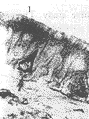
, 百拇医药
图1 兔气管 Fig.1 The trachea of rabbit(↑)示锥形嗜银细胞。双重银染改良法×268;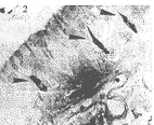
图2 豚鼠气管 Fig.2 The trachea of guinea pig(↑)示卵圆形、梭形嗜银细胞。双重银染改良法×134;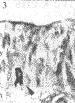
图3 豚鼠气管 Fig.3 The trachea of guinea pig(↑)示有突起的嗜银细胞。双重银染改良法×134;
2.2 5-HT-IR细胞 用PAP法显示的5-HT-IR细胞背景清晰,胞质内含棕色或棕黄色颗粒,对照切片均为阴性。狗和大鼠气管上皮内未见到5-HT-IR细胞。家兔、豚鼠及小鼠气管内5-HT-IR细胞均较少,呈单个分散在上皮细胞之间,细胞为柱状、梭形、锥形等。柱状细胞的顶端达腔面,为开放型细胞(图5),梭形、锥形细胞顶端一般不达腔面,为闭合型细胞(图4,6)。5-HT-IR细胞的密度,家兔大于豚鼠和小鼠。
, http://www.100md.com
图4 小鼠气管 Fig.4 The trachea of mouse(↑)示梭形5-HT-IR细胞。免疫组化PAP法×268;
图5 兔气管 Fig.5 The trachea of rabbit(↑)示达腔面的5-HT-IR细胞。免疫组化PAP法×268;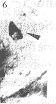
图6 小鼠气管 Fig.6 The trachea of mouse(↑)示锥形5-HT-IR细胞。免疫组化PAP法×268
2.3 相邻切片观察未发现嗜银颗粒和5-HT共存于气管上皮内同一细胞。
3 讨论
, http://www.100md.com
呼吸道粘膜上皮内广泛存在的内分泌细胞,在肺报道较多[8~10],气管报道甚少。本实验用双重银染法显示的结果表明,5种动物气管上皮内均有散在的嗜银细胞,此结果与Hage等[2]和Cutz等[3]用Grimelius银染法对人和家兔气管的观察结果相一致。用免疫组织化学方法特异地显示了家兔、豚鼠及小鼠气管粘膜上皮内有5-HT-IR细胞,此结果与Ericson等[4]和Richard等[5]用Falck荧光胺法分别发现小鼠和家兔气管粘膜内有显示5-HT存在的黄绿色荧光的结果相似。从而证实了气管上皮的内分泌细胞含有5-HT。嗜银细胞的密度在家兔较多而狗、大鼠较少以及5-HT-IR细胞仅见于家兔、豚鼠和小鼠的结果,可能与动物种属间差异有关。
气管上皮的嗜银细胞和5-HT-IR细胞可分为开放型和闭合型。本实验还观察到嗜银细胞尚有旁突伸至相邻细胞,故气管内分泌细胞也具有旁分泌功能的形态学依据。本文结果表明了气管内分泌细胞具有APUD细胞的形态特征。这类细胞可能通过腔分泌、旁分泌或经血液循环参与呼吸道血管平滑肌收缩和腺体的分泌[11]。近年来对肺的内分泌细胞的研究已经证明,肺内有分泌蛙皮素、胃泌素、降钙素和5-HT等多种内分泌细胞[10]。至于气管内是否存在分泌其他肽类物质的内分泌细胞,尚待进一步研究证实。
, http://www.100md.com
参考文献
1,Pearse A G E.The cytochemistry and ultrastructure of polypeptide hormone-producing cells of the APUD series and the embryologic, physiologic and pathologic implication of the concept. J histochem cytochem, 1969,17:303
2,Hage E.Endocrine like cells of the pulmonary epthelium of the human adult lung. Cell Tiss Res, 1977,178:39
3,Cutz E. Ultrastructure and fluorescene histochemistry of endocrine (APUD-type)cells in trachea of human and various animal species, Cell Tiss Res, 1975, 158:425
, 百拇医药
4,Ericson L E. Fluorescence and electron microscopy of amine-stroring enterochromaffin-like cells in trealeal epthelium of mouse. Z.Zellforsch, 1972,124:532-545
5,Richard D. Morphology histochemistry and distribution of serotin-containing cells in tracheal epthelium of adult rabbit. Anat Res, 1981,199:23
6,何素云,许射阜.显示APUD细胞的新方法-双重银染改良法.贵阳医学院报,1985,10:158
7,Sternberger LA. Immunocytochemistry. New York:John Wiley and Sons, 1979.104-169
, http://www.100md.com
8,Hage E. Histoehemistry and fine structure of endocrine cells in fatal lungs of the rabbit, mouse and guinea pig. Cell Tiss Res, 1974,149:513
9,Hage E. Electron microscopic identification of endocrine cells in the bronchial epithelium of human futuses.Acta Pathol Microbiol Immunol Scandi [A], 1972, 80:143
10,Gosney JR. Neuroendocrine cell population in normal human lungs:A quantitative study.Thorax,1988, 48:878
11,成令忠主编.组织学.北京:人民卫生出版社,1993.1242~1280
(1999―03―24收稿,1999―05―26修回), http://www.100md.com