小鼠胚着床前线粒体的分布和超微结构变化*
作者:韩贻仁 赵 晖
单位:山东大学生命科学学院,济南 250100
关键词:线粒体;着床前胚;小鼠
解剖学报980318 摘 要 为了解小鼠胚着床前细胞中线粒体的分布和超微结构的变化规律,观察了2细胞胚、4细胞胚、8细胞胚、桑椹胚、早期囊胚和晚期囊胚。2细胞期和桑椹胚期,线粒体绕胞核密集,在挤紧的8细胞胚中,线粒体在细胞接触面处的胞质边缘密集。 囊胚期, 滋养层细胞的线粒体在胞核周围较宽的区域中分布。囊胚经罗丹明123染色后, 在荧光显微镜下显现为颗粒状。挤紧的8细胞胚,经秋水仙素(10mg/L)处理后, 线粒体在胞质中变为弥散分布。 电镜下可见从受精卵至4细胞期,线粒体呈球形、无嵴、中央有低电子密度区。8细胞期,线粒体呈球形或圆形,内部出现嵴。桑椹期至囊胚期, 线粒体变长,呈杆状,有横嵴。
哺乳动物着床前胚在发育过程中发生一系列生理和形态变化,如分裂球间的挤紧作用(compaction),细胞间连接的形成[1]、囊胚腔的产生、以及内细胞团和滋养层2个细胞系分化[2]等。线粒体是为细胞各种活动提供能源物质——ATP的细胞器。Blerkom[3]发现,小鼠卵母细胞在成熟过程中,线粒体的分布发生了2次转移,即线粒体在一定时期向某些区域暂时密集, 为细胞的局部活动提供较多的ATP。 我们在以前[4]已报道了小鼠受精卵在第1次卵裂过程中线粒体的分布变化。关于小鼠胚着床前发育过程中线粒体分布的变化规律尚未见报道, 本研究对此作了观察, 同时对线粒体超微结构的变化进行了分析。
, http://www.100md.com
材料和方法
1.动物
雌性成体昆明小鼠。体重25~30g,按雌、雄比3∶1自然交配受孕。受孕时间由发现阴栓当日的0时算起。
2.胚胎的收集
在受孕32h\,46h\,57h\,65h\,70h\,82h和104h,断头杀死雌鼠,用Whitten液由输卵管或子宫分别冲取2细胞胚、4细胞胚、8细胞胚、桑椹胚、早期囊胚和晚期囊胚。冲胚的方法参见文献[5]。 在冲胚前,向盛有1ml Whitten液的试管中充入5%CO2\,5%O2和90%N2混合气体(北京综合仪器厂产)15s,调培养液pH值至7.2~7.4[6]。
3.培养液的配制
按Whitten配方配制供着床前胚体外生长的培养液[7], 用孔径0.3μm的微孔滤膜过滤除菌。
, 百拇医药
向Whitten培养液中加入罗丹明123(rhodamine 123, Sigma产品)或秋水仙素(Fluka产品)分别配制成罗丹123培养液(10mg/L)和秋水仙素培养液(10mg/L)。
4.荧光染色和透射电镜观察
将冲取出的不同阶段的胚分别取5~10枚放入盛有1ml罗丹明123培养液的小试管中, 培养10min, 再用Whitten液换洗3次,置载片上,于Olympus BH-2荧光显微镜下观察线粒体的分布。
秋水仙素实验组是先将5~10枚挤紧的8细胞胚放入盛有1ml秋水仙素培养液的试管中培养1h, 再用Whitten培养液换洗1次,放入罗丹明123培养液中染色、观察。
以上处理过程,详见文献[4]。
取出各期胚后, 按常规程序制备电镜标本,Epon 812包埋,LKB切片,100CX透射电镜观察。
, 百拇医药
结 果
1.不同发育阶段胚中线粒体的分布形式
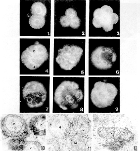
线粒体在胚细胞中的分布形式具有阶段性特征。在2细胞期,线粒体(M)明显围核集中分布(图1)。4细胞期,M在核周仍较密集(图2)。8细胞早期,M在细胞质中分布较均匀,绕核集中现象不明显(图3);8细胞胚发生挤紧之后,M除在胞核周围密集外,尚在相邻细胞接触面的质膜下方明显密集(图4)。至桑椹胚,M在胞核周围区域集中分布(图5)。在囊胚早期和中期,滋养层细胞的M绕胞核集中,而在扩张囊胚中,M在胞核周围较宽的区域中密集(图6,7)。囊胚期,荧光显示的M轮廓呈较清晰的颗粒状(图8)。
2.秋水仙素对线粒体分布的影响
挤紧的8细胞胚经秋水仙素处理后,M在细胞质中弥散分布,在相邻细胞接触面处的质膜下方,M集中现象消退(图9)。
, 百拇医药
3.不同发育阶段胚线粒体的超微结构
着床前胚从受精卵至4细胞期,M呈球形或椭圆形,无嵴,中央有低电子密度区(图10)。至8细胞期,M仍呈球形或椭圆形,内部出现了嵴(图11)。 在桑椹胚至囊胚,M变为长形,横嵴明显(图12)。
讨 论
线粒体是真核细胞中的一种重要细胞器,为细胞的多种生理活动提供能源。小鼠卵细胞在卵原细胞阶段,M呈长条形,有横嵴。在初级卵母细胞生长期,M由长条形逐渐转变为球形或椭圆形,体积减小,但数量增加[7]。M的小型化有利于在体积较大的胚胎细胞中移动,易于向需能部位汇集。在受精卵中M均匀分布,只在原核周围略密[4]。从受精卵到囊胚的发育过程中,M集中的部位有3次明显的移动。第1次发生在受精卵至2细胞期,M向胞核周围密集;第2次发生在8细胞期,M由8细胞期的均匀分布到胚胎挤紧后向细胞接触面胞质集中;第3次是在桑椹胚期,M又向核周围区域密集。仓鼠胚着床前,M的分布形式亦发生类似动态变化[8]。
, http://www.100md.com
M的分布形式和结构的变化与受精卵发育中生理代谢和细胞结构变化存在对应关系。1细胞期,核活动较弱,蛋白质的合成主要依靠母体mRNA进行[9],这时M在胞质中均匀分布。2细胞期,胚胎基因组开启表达,核活动增强,M的围核集中可能反映了核与细胞质物质交换的增加。8细胞期ATP合成量增加[10],在8细胞后期发生了重要的挤紧变化,分裂球间的接触面扩大,细胞变成顶宽底窄的锥形,细胞器在胞质中分布不均,产生了极性[11]。在相邻细胞顶端相互接触面处形成了紧密连接和间隙连接[12],细胞间相互联系更加密切,这些变化主要发生在细胞接触面处,M在此处胞质中密集,显然有利于为此处变化提供ATP。桑椹胚期,胚胎细胞分化为滋养层和内细胞团2个细胞系。囊胚期形成囊胚腔,滋养层细胞的主要活动是吸收胚外物质,穿过滋养层细胞进入囊胚腔的葡萄糖量大为增加,腔的体积扩大[13],M在核周围较宽的区域内密集适合于物质的穿胞运输。挤紧的胚胎经过秋水仙素处理后,M的分布失去了局部密集的特征, 说明M的分布形式可能与微管的作用有关,这与我们前文报道的结果一致[4]。
, http://www.100md.com
我们的实验还表明,在小鼠着床前胚发育过程中,M本身的结构亦在逐渐发生变化。在1~4细胞期,M是小球形。在8细胞期,M呈椭圆形,内部出现嵴。在桑椹胚和囊胚期,M变为长杆状,内部有横嵴,近似于成体细胞中的M形态。这种结构变化反映出M合成ATP的能力加强和细胞活动对ATP需求量的增加[14,15]。由此推想,囊胚经罗丹明123染色后,M呈现出较清晰的颗粒状轮廓,可能是其M结构分化成熟的反映。
我们的研究表明,着床前胚发育过程中,M的分布处于动态变化之中,其分布形式具有阶段性特征,与细胞对ATP的局部需求相适应。卵母细胞和着床前胚M的小形化有利于M的移动。
收稿 1997-02 修回 1997-07
* 山东省科学技术委员会资助课题(No.179),鲁科计字(1994)
参考文献
, 百拇医药
[1]Ducibella T.Surface changes of developing trophoblast cell. In:Johnson MH(ed). Development in Mammals.vol 1. Amsterdam:Elsevier/North-Holland Biomedical Press, 1977:5-30
[2]Pedersen RA. Potency, lineage, and allocation in preimplantation mouse embryo. In:Approaches to Mammalian Embryonic Development. Cambridge:Cambridge University Press, 1988:3-33
[3]Van Blerkom J, Runner MN. Mitochondrial reorganization during resumption of arrest meiosis in the mouse oocyte. Am J Anat, 1984, 171(3):335
, http://www.100md.com
[4]韩贻仁,张振玲.小鼠第一次卵裂周期中线粒体分布的变化.解剖学报,1994,25(31):286
[5]韩贻仁,蓝厚珍.昆明鼠早期胚胎发育的初步观察。山东大学学报(自然科学版),1979,12(3):107
[6]韩贻仁.国产试剂配制的培养液对小鼠早期胚胎培养效果的研究.山东大学学报(自然科学版),1989,24(3):128
[7]Wassarman PM, Josefowicz WJ. Oocyte development in the mouse:An ultrastructural comparision of oocytes isolated at various stages of growth and meiotic competance. J Morphol, 1978, 156(1):209
, 百拇医药 [8]Barnet DK, Kimura JL, Bavister BD, et al. Translocation of active mitochondria during hamster preimplantation embryo development studied by confocal laser scanning microscopy. Dev Dyn, 1996,205(3):286
[9]Clegg KB, Piko L. Poly(A) length, cytoplasmic adenylation and synthesis of poly(A) +RNA in early mouse embryos. Dev Biol, 1983,95(2):331
[10]Ginsberg L, Hillman N. ATP metabolism in cleavage-staged mouse embryos. J Embryol Exp Morphol, 1973, 30(1):262
, http://www.100md.com
[11]Johnson MH, Ziomek CA. Induction of polarity in mouse 8-cell blastomere:specificity, geometry and stability. J Cell Biol, 1981, 91(1):303
[12]Ducibella T, Albertini DF, Anderson E, et al. The preimplantation mammalian embryo:characterizaton of intercellular junctions and their appearance during development. Dev Biol, 1975, 45(1):231
[13]Gardner HG, Kaye PL. Characterizaton of glucose transport in preimplantation mouse embryos. Reprod Fertil Dev, 1995,7(1):41
, 百拇医药
[14]Mills RM, Brinster RL. Oxygen consumption of preimplantation mouse embryos. Exp Cell Res, 1967, 47(1/2):337
[15]Leese HJ. Metabolism of the preimplantation mammalian embryos. In:Milligan SR(ed). Oxford Reviews of Reproductive Biology, vol 13. New York:Oxford University Press, 1991:35-72
THE CHANGES OF THE DISTRIBUTION PATTERNS AND
ULTRASTRUCTURE OF MITOCHONDRIA DURING
, 百拇医药
PREIMPLANTATION DEVELOPMENT OF
MOUSE EMBRYOS
Han Yiren, Zhao Hui
(School of Life Science, Shandong University, Ji'nan)
The ultrastructure and distribution patterns of mitochondria in blastomeres are successively changing during preimplantation development of mouse embryo, and present specific features at various stages. In the development of early preimplantation embryo there are three major translocations of mitochondria in cytoplasm of blastomere. The first translocation is between 1-cell and 2-cell stages, which results in perinuclear concentration of mitochondria.The second is at 8-cell stage, during compaction of embryo the distribution patterns of mitochondria are from even dispersion to aggregate in the margin area of cytoplasm along the surfaces of opposite blastomeres. The third is at morula stage, in which mitochondria reaggregate in a broader region around nucleus. At blastula stage the mitochondria of trophoblast still reveal a perinuclear arrangement in a broader area of cytoplasm, and they are viewed as clear granules under fluorescence microscope. According to the phenomena of mitochondrial dispersion throughout the blastomeres cytoplasm of compacted 8-cell embryo which was exposed to colchicine, we suggest that the microtubules probably play a key role in maintenance of the distribution patterns of mitochondria in blastomeres. Ultrastructures indicate that at different stages of embryo, the appearance of mitochondria has different features. In the 1 to 4 cell embryos mitochondra are spheroidal in shape with a low electron density area in the center, and there are no cristae in it. In the blastomeres of 8 cell embryos the mitochondrion has a ovoid shape and contains a few cristae.The majority of the mitochondria of morula and blastula are elongated in structures with transverse cristae within them.Thus, the transmission electron microscopic and fluorescence microscopic observations demonstrated that during development of preimplantation embryos the mitochondria develop from a simple spherical appearance to more mature form which would be convenient to provide increasing amounts of ATP for further developmental events.
, 百拇医药
KEY WORDS Mitochondrion; Preimplantation embryo; Mouse
△School of Life Science, Shandong University, Ji'nan 250100,China
《解剖学报》编辑委员会
顾 问 杨 进
主 编 章静波
副主编 徐群渊 孙品伟 许增禄 于恩华
编 委 贲长恩 蔡文琴 陈尔瑜 董新文 高英茂 郭仁强 郭畹华 韩永坚 何素云
胡人义 姜均本 鞠 躬 刘 斌 饶志仁 石玉秀 苏慧慈 田竟生 王国英
王云祥 吴晋宝 吴良芳 吴新智 张世馥 张为龙 周长满 周明华 朱长庚
宗书东 左焕琛
(以姓氏汉语拼音为序), 百拇医药
单位:山东大学生命科学学院,济南 250100
关键词:线粒体;着床前胚;小鼠
解剖学报980318 摘 要 为了解小鼠胚着床前细胞中线粒体的分布和超微结构的变化规律,观察了2细胞胚、4细胞胚、8细胞胚、桑椹胚、早期囊胚和晚期囊胚。2细胞期和桑椹胚期,线粒体绕胞核密集,在挤紧的8细胞胚中,线粒体在细胞接触面处的胞质边缘密集。 囊胚期, 滋养层细胞的线粒体在胞核周围较宽的区域中分布。囊胚经罗丹明123染色后, 在荧光显微镜下显现为颗粒状。挤紧的8细胞胚,经秋水仙素(10mg/L)处理后, 线粒体在胞质中变为弥散分布。 电镜下可见从受精卵至4细胞期,线粒体呈球形、无嵴、中央有低电子密度区。8细胞期,线粒体呈球形或圆形,内部出现嵴。桑椹期至囊胚期, 线粒体变长,呈杆状,有横嵴。
哺乳动物着床前胚在发育过程中发生一系列生理和形态变化,如分裂球间的挤紧作用(compaction),细胞间连接的形成[1]、囊胚腔的产生、以及内细胞团和滋养层2个细胞系分化[2]等。线粒体是为细胞各种活动提供能源物质——ATP的细胞器。Blerkom[3]发现,小鼠卵母细胞在成熟过程中,线粒体的分布发生了2次转移,即线粒体在一定时期向某些区域暂时密集, 为细胞的局部活动提供较多的ATP。 我们在以前[4]已报道了小鼠受精卵在第1次卵裂过程中线粒体的分布变化。关于小鼠胚着床前发育过程中线粒体分布的变化规律尚未见报道, 本研究对此作了观察, 同时对线粒体超微结构的变化进行了分析。
, http://www.100md.com
材料和方法
1.动物
雌性成体昆明小鼠。体重25~30g,按雌、雄比3∶1自然交配受孕。受孕时间由发现阴栓当日的0时算起。
2.胚胎的收集
在受孕32h\,46h\,57h\,65h\,70h\,82h和104h,断头杀死雌鼠,用Whitten液由输卵管或子宫分别冲取2细胞胚、4细胞胚、8细胞胚、桑椹胚、早期囊胚和晚期囊胚。冲胚的方法参见文献[5]。 在冲胚前,向盛有1ml Whitten液的试管中充入5%CO2\,5%O2和90%N2混合气体(北京综合仪器厂产)15s,调培养液pH值至7.2~7.4[6]。
3.培养液的配制
按Whitten配方配制供着床前胚体外生长的培养液[7], 用孔径0.3μm的微孔滤膜过滤除菌。
, 百拇医药
向Whitten培养液中加入罗丹明123(rhodamine 123, Sigma产品)或秋水仙素(Fluka产品)分别配制成罗丹123培养液(10mg/L)和秋水仙素培养液(10mg/L)。
4.荧光染色和透射电镜观察
将冲取出的不同阶段的胚分别取5~10枚放入盛有1ml罗丹明123培养液的小试管中, 培养10min, 再用Whitten液换洗3次,置载片上,于Olympus BH-2荧光显微镜下观察线粒体的分布。
秋水仙素实验组是先将5~10枚挤紧的8细胞胚放入盛有1ml秋水仙素培养液的试管中培养1h, 再用Whitten培养液换洗1次,放入罗丹明123培养液中染色、观察。
以上处理过程,详见文献[4]。
取出各期胚后, 按常规程序制备电镜标本,Epon 812包埋,LKB切片,100CX透射电镜观察。

, 百拇医药
结 果
1.不同发育阶段胚中线粒体的分布形式
线粒体在胚细胞中的分布形式具有阶段性特征。在2细胞期,线粒体(M)明显围核集中分布(图1)。4细胞期,M在核周仍较密集(图2)。8细胞早期,M在细胞质中分布较均匀,绕核集中现象不明显(图3);8细胞胚发生挤紧之后,M除在胞核周围密集外,尚在相邻细胞接触面的质膜下方明显密集(图4)。至桑椹胚,M在胞核周围区域集中分布(图5)。在囊胚早期和中期,滋养层细胞的M绕胞核集中,而在扩张囊胚中,M在胞核周围较宽的区域中密集(图6,7)。囊胚期,荧光显示的M轮廓呈较清晰的颗粒状(图8)。
2.秋水仙素对线粒体分布的影响
挤紧的8细胞胚经秋水仙素处理后,M在细胞质中弥散分布,在相邻细胞接触面处的质膜下方,M集中现象消退(图9)。
, 百拇医药
3.不同发育阶段胚线粒体的超微结构
着床前胚从受精卵至4细胞期,M呈球形或椭圆形,无嵴,中央有低电子密度区(图10)。至8细胞期,M仍呈球形或椭圆形,内部出现了嵴(图11)。 在桑椹胚至囊胚,M变为长形,横嵴明显(图12)。
讨 论
线粒体是真核细胞中的一种重要细胞器,为细胞的多种生理活动提供能源。小鼠卵细胞在卵原细胞阶段,M呈长条形,有横嵴。在初级卵母细胞生长期,M由长条形逐渐转变为球形或椭圆形,体积减小,但数量增加[7]。M的小型化有利于在体积较大的胚胎细胞中移动,易于向需能部位汇集。在受精卵中M均匀分布,只在原核周围略密[4]。从受精卵到囊胚的发育过程中,M集中的部位有3次明显的移动。第1次发生在受精卵至2细胞期,M向胞核周围密集;第2次发生在8细胞期,M由8细胞期的均匀分布到胚胎挤紧后向细胞接触面胞质集中;第3次是在桑椹胚期,M又向核周围区域密集。仓鼠胚着床前,M的分布形式亦发生类似动态变化[8]。
, http://www.100md.com
M的分布形式和结构的变化与受精卵发育中生理代谢和细胞结构变化存在对应关系。1细胞期,核活动较弱,蛋白质的合成主要依靠母体mRNA进行[9],这时M在胞质中均匀分布。2细胞期,胚胎基因组开启表达,核活动增强,M的围核集中可能反映了核与细胞质物质交换的增加。8细胞期ATP合成量增加[10],在8细胞后期发生了重要的挤紧变化,分裂球间的接触面扩大,细胞变成顶宽底窄的锥形,细胞器在胞质中分布不均,产生了极性[11]。在相邻细胞顶端相互接触面处形成了紧密连接和间隙连接[12],细胞间相互联系更加密切,这些变化主要发生在细胞接触面处,M在此处胞质中密集,显然有利于为此处变化提供ATP。桑椹胚期,胚胎细胞分化为滋养层和内细胞团2个细胞系。囊胚期形成囊胚腔,滋养层细胞的主要活动是吸收胚外物质,穿过滋养层细胞进入囊胚腔的葡萄糖量大为增加,腔的体积扩大[13],M在核周围较宽的区域内密集适合于物质的穿胞运输。挤紧的胚胎经过秋水仙素处理后,M的分布失去了局部密集的特征, 说明M的分布形式可能与微管的作用有关,这与我们前文报道的结果一致[4]。
, http://www.100md.com
我们的实验还表明,在小鼠着床前胚发育过程中,M本身的结构亦在逐渐发生变化。在1~4细胞期,M是小球形。在8细胞期,M呈椭圆形,内部出现嵴。在桑椹胚和囊胚期,M变为长杆状,内部有横嵴,近似于成体细胞中的M形态。这种结构变化反映出M合成ATP的能力加强和细胞活动对ATP需求量的增加[14,15]。由此推想,囊胚经罗丹明123染色后,M呈现出较清晰的颗粒状轮廓,可能是其M结构分化成熟的反映。
我们的研究表明,着床前胚发育过程中,M的分布处于动态变化之中,其分布形式具有阶段性特征,与细胞对ATP的局部需求相适应。卵母细胞和着床前胚M的小形化有利于M的移动。
收稿 1997-02 修回 1997-07
* 山东省科学技术委员会资助课题(No.179),鲁科计字(1994)
参考文献
, 百拇医药
[1]Ducibella T.Surface changes of developing trophoblast cell. In:Johnson MH(ed). Development in Mammals.vol 1. Amsterdam:Elsevier/North-Holland Biomedical Press, 1977:5-30
[2]Pedersen RA. Potency, lineage, and allocation in preimplantation mouse embryo. In:Approaches to Mammalian Embryonic Development. Cambridge:Cambridge University Press, 1988:3-33
[3]Van Blerkom J, Runner MN. Mitochondrial reorganization during resumption of arrest meiosis in the mouse oocyte. Am J Anat, 1984, 171(3):335
, http://www.100md.com
[4]韩贻仁,张振玲.小鼠第一次卵裂周期中线粒体分布的变化.解剖学报,1994,25(31):286
[5]韩贻仁,蓝厚珍.昆明鼠早期胚胎发育的初步观察。山东大学学报(自然科学版),1979,12(3):107
[6]韩贻仁.国产试剂配制的培养液对小鼠早期胚胎培养效果的研究.山东大学学报(自然科学版),1989,24(3):128
[7]Wassarman PM, Josefowicz WJ. Oocyte development in the mouse:An ultrastructural comparision of oocytes isolated at various stages of growth and meiotic competance. J Morphol, 1978, 156(1):209
, 百拇医药 [8]Barnet DK, Kimura JL, Bavister BD, et al. Translocation of active mitochondria during hamster preimplantation embryo development studied by confocal laser scanning microscopy. Dev Dyn, 1996,205(3):286
[9]Clegg KB, Piko L. Poly(A) length, cytoplasmic adenylation and synthesis of poly(A) +RNA in early mouse embryos. Dev Biol, 1983,95(2):331
[10]Ginsberg L, Hillman N. ATP metabolism in cleavage-staged mouse embryos. J Embryol Exp Morphol, 1973, 30(1):262
, http://www.100md.com
[11]Johnson MH, Ziomek CA. Induction of polarity in mouse 8-cell blastomere:specificity, geometry and stability. J Cell Biol, 1981, 91(1):303
[12]Ducibella T, Albertini DF, Anderson E, et al. The preimplantation mammalian embryo:characterizaton of intercellular junctions and their appearance during development. Dev Biol, 1975, 45(1):231
[13]Gardner HG, Kaye PL. Characterizaton of glucose transport in preimplantation mouse embryos. Reprod Fertil Dev, 1995,7(1):41
, 百拇医药
[14]Mills RM, Brinster RL. Oxygen consumption of preimplantation mouse embryos. Exp Cell Res, 1967, 47(1/2):337
[15]Leese HJ. Metabolism of the preimplantation mammalian embryos. In:Milligan SR(ed). Oxford Reviews of Reproductive Biology, vol 13. New York:Oxford University Press, 1991:35-72
THE CHANGES OF THE DISTRIBUTION PATTERNS AND
ULTRASTRUCTURE OF MITOCHONDRIA DURING
, 百拇医药
PREIMPLANTATION DEVELOPMENT OF
MOUSE EMBRYOS
Han Yiren, Zhao Hui
(School of Life Science, Shandong University, Ji'nan)
The ultrastructure and distribution patterns of mitochondria in blastomeres are successively changing during preimplantation development of mouse embryo, and present specific features at various stages. In the development of early preimplantation embryo there are three major translocations of mitochondria in cytoplasm of blastomere. The first translocation is between 1-cell and 2-cell stages, which results in perinuclear concentration of mitochondria.The second is at 8-cell stage, during compaction of embryo the distribution patterns of mitochondria are from even dispersion to aggregate in the margin area of cytoplasm along the surfaces of opposite blastomeres. The third is at morula stage, in which mitochondria reaggregate in a broader region around nucleus. At blastula stage the mitochondria of trophoblast still reveal a perinuclear arrangement in a broader area of cytoplasm, and they are viewed as clear granules under fluorescence microscope. According to the phenomena of mitochondrial dispersion throughout the blastomeres cytoplasm of compacted 8-cell embryo which was exposed to colchicine, we suggest that the microtubules probably play a key role in maintenance of the distribution patterns of mitochondria in blastomeres. Ultrastructures indicate that at different stages of embryo, the appearance of mitochondria has different features. In the 1 to 4 cell embryos mitochondra are spheroidal in shape with a low electron density area in the center, and there are no cristae in it. In the blastomeres of 8 cell embryos the mitochondrion has a ovoid shape and contains a few cristae.The majority of the mitochondria of morula and blastula are elongated in structures with transverse cristae within them.Thus, the transmission electron microscopic and fluorescence microscopic observations demonstrated that during development of preimplantation embryos the mitochondria develop from a simple spherical appearance to more mature form which would be convenient to provide increasing amounts of ATP for further developmental events.
, 百拇医药
KEY WORDS Mitochondrion; Preimplantation embryo; Mouse
△School of Life Science, Shandong University, Ji'nan 250100,China
《解剖学报》编辑委员会
顾 问 杨 进
主 编 章静波
副主编 徐群渊 孙品伟 许增禄 于恩华
编 委 贲长恩 蔡文琴 陈尔瑜 董新文 高英茂 郭仁强 郭畹华 韩永坚 何素云
胡人义 姜均本 鞠 躬 刘 斌 饶志仁 石玉秀 苏慧慈 田竟生 王国英
王云祥 吴晋宝 吴良芳 吴新智 张世馥 张为龙 周长满 周明华 朱长庚
宗书东 左焕琛
(以姓氏汉语拼音为序), 百拇医药