CD57+自然杀伤细胞在妊娠早期小鼠子宫壁内的分布*
作者:钟秀会 周占祥 孙秉贵 邓泽沛 刘济五
单位:中国农业大学动物医学院解剖学组织胚胎学教研室,北京 100094
关键词:NK细胞;妊娠早期;妊娠失败;小鼠
解剖学报990424 【摘要】 目的 探索CD57+自然杀伤细胞(NK细胞,natural killer cells)在小鼠妊娠早期子宫壁内的意义。方法 用小鼠抗人NK细胞CD57单克隆抗体,对未经产小鼠妊娠早期子宫壁内NK细胞进行免疫组织化学标记。结果 孕0d,小鼠子宫壁内见少量CD57+细胞,孕2d时NK细胞数量剧增,孕4d时稍下降。孕6d、孕8d时数量更少(低于0d值)。孕6d和8d均出现妊娠失败个体,其子宫壁内NK细胞很多,显著高于正常妊娠6d和8d小鼠的数量(P<0.01)。从分布区域看,胚胎着床(4.5d)前,NK细胞多位于子宫肌层,胚胎着床后仅存在于着床点以外的子宫内膜和肌层。在妊娠失败个体,NK细胞广泛存在于子宫内膜和肌层中。 结论 小鼠交配后子宫NK细胞增多与子宫炎性反应有关。着床期以后子宫NK细胞增多与妊娠失败有一定的相关性。
, 百拇医药
DISTRIBUTION OF CD57+ NATURAL KILLER CELLS IN
MOUSE UTERUS DURING EARLY PREGNANCY
Zhong Xiuhui, Zhou Zhanxiang△,Sun Binggui, Deng Zepei, Liu Jiwu
(Department of Animal Anatomy and Embryology, College of Veterinary Medicine, China Agricultural University, Beijing)
【Abstract】 Objective To realize the significance of CD57+ natural killer cells in mouse uterus during early pregnancy. Methods Using a mouse-anti-human CD57 monoclonal antibody to localize NK cells in uterus of virgin mice. Results There are a few CD57+ cells in the uterus on day 0 of pregnancy. The NK cells are elevated sharply in number on day 2 and become less on day 4. On days 6 and 8 of normal pregnancies, the cell numbers are much decreased(lower than the value of day 0). At this time, pregnancy failure was found in some individuals, in which the CD57+ cells occur in great numbers in the uteri. In reference to distribution, the NK cells are usually found in the myometrium prior to implantation (day 4.5). NK cells only occur in the undecidualized endometrium or myometrium post implantation in normal pregnancies. In pregnancy failure,however, the cells are evenly distributed in whole uterus.Conclusion The NK cells in mouse uterus increase after mating, implying the participation of NK cells in the inflammatory response of the uterus to mating and the elevation of NK cells in the uterus after the time of implantation is associated with pregnancy failure.
, 百拇医药
【Key words】 Natural killer cells; Early pregnancy; Pregnancy failure; Mouse
哺乳动物妊娠过程中,胚胎着床成功与否受母体免疫环境的直接影响,子宫NK细胞是其中重要的免疫组分。近年来,以鼠类作为动物模型,对NK细胞在妊娠子宫中的分布、增殖以及功能做了较详尽的研究[1],但对NK细胞在胚胎着床过程中的意义仍不清楚。
研究小鼠NK细胞国外常用的抗体有NK1.1、Asialo-GM1,以及近年来研制的泛NK细胞单克隆抗体DX5等。对于小鼠抗人CD57+单克隆抗体,虽有技术资料(美国NeoMarkers公司)介绍能够识别小鼠、大鼠、仓鼠和鸡的NK细胞,但迄今国内外未见试验研究报道。本试验用CD57单抗研究了NK细胞在妊娠早期小鼠子宫壁内的分布,以探索CD57+细胞在妊娠早期小鼠子宫壁内存在的意义。
, http://www.100md.com
材料和方法
1.动物及取材
8周龄未经产昆明种小鼠。常规饲养,自由采食饮水。每日光照12h。经适应1周后阴道涂片法作发情检查,发情者与公鼠(1∶1)同笼过夜,次日早晨检查有阴栓者为孕0d。取材共分7组,每组小鼠5只。孕0、2、4d各1组。孕6d、孕8d各取2组。6A、8A为妊娠正常组。子宫角外观有串珠状隆起,胚胎个数清晰可见。后经切片组织学检查胚胎发育正常。6B、8B为胚胎丢失组,子宫角比未孕子宫角粗大些,未见明显胚胎隆起。切片中见到蜕膜化子宫内膜未完全复旧。
颈椎脱臼法处死孕鼠,剖腹取出子宫,波恩液固定48h,梯度酒精脱水,Paraplast(美国Sigma公司)包埋。滑动切片机纵切(厚6μm)。
2.免疫组织化学染色
切片脱蜡入水后,经H2O2封闭,抗原热修复,进行免疫组织化学染色。主要程序如下:10%犊牛血清封闭,滴加CD57单克隆抗体(北京邦定生物医学公司),4℃过夜,生物素化羊抗鼠IgM(美国Vector公司)37℃孵育30min,HRP标记链霉亲和素(life technologies,Inc.)37℃孵育30min,每步骤之间用0.01mol/L PBS(pH7.4)冲洗3次。0.03%DAB显色5~10min,常规脱水透明,封片。
, 百拇医药
3.细胞计数
40倍物镜下观察,每样品取20个视野,统计阳性细胞总数,计算组内个体间的平均值。t检验法计算组间差异显著性。
结果和讨论
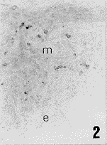
孕0d,子宫壁内见少量CD57+细胞,散在于肌层(图1)。孕2d时,CD57+细胞增多,从0d时的16.3个增加到62.3个(P<0.01),仍分布于子宫肌层(图2)。有些个体可见CD57+细胞成簇存在于子宫内膜和内环肌之间。孕2d时子宫NK细胞增多,可能与交配引起的子宫炎性反应有关,说明NK细胞在胚胎着床前可能起着抗感染作用。
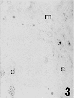
孕4d时,子宫内膜蜕膜化,进入胚胎着床期(4.5d完成着床)。此时子宫壁内CD57+细胞比孕2d时减少。但仍高于0d(P<0.01)。细胞存在于着床点以外未蜕膜化的子宫内膜和肌层中(图3)。这显示,胚胎植入不依赖NK细胞的存在。
, 百拇医药
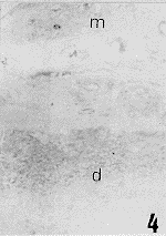
孕6d和孕8d剖杀的小鼠出现两种情况,一种是妊娠正常者,子宫角外观有串珠状隆起,内含发育正常的胚胎。此时子宫壁内CD57+细胞很少,位于胚胎着床点以外的肌层(图4)。说明CD57+细胞不是胚胎存活和生长所需要的细胞成分[2]。
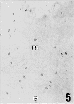
孕6d和孕8d时的第2种情况是出现妊娠失败个体。子宫角内没有胚胎,看不到串珠状隆起,仅比未孕子宫角粗大些。子宫内CD57+细胞很多,遍布于子宫内膜和肌层中(图5)。表明子宫壁内NK细胞增多与妊娠失败有密切关系。虽然本试验结果不能直接证明,子宫壁内NK细胞增多参与或直接引起妊娠失败的发生,但从前人研究结果来看,抑制NK细胞活性能显著降低孕鼠胚胎吸收率[3],激活NK细胞活性则增加胚胎吸收率[4],说明NK细胞增多是诱发胚胎吸收的原因。加拿大学者Gendron和Baines[5]也发现,子宫壁内NK细胞增多是小鼠发生早期自发性流产的诱因。由此我们有理由推论,本试验观察到的小鼠孕6d、孕8d时的妊娠失败,与子宫壁内CD57+NK的细胞增多有关。
, http://www.100md.com
CD57+细胞在不同妊娠日龄小鼠子宫壁内的数量分布及组间差异性比较结果见附表。
附表 小鼠妊娠早期子宫内CD57+细胞的分布
Table CD57+ cells in mouse uterus in early pregnancy 妊娠日龄
day of pregnancy
0d
2d
4d
6d
8d
, http://www.100md.com 正常妊娠
normal pregnancy
16.3±
2.4
62.3±
7.6a,b
33±
5.2a,b
9.7±
1.3
7.3±
1.3
, http://www.100md.com
妊娠失败
pregnancy failure
65.7±
7.2c
64.6±
4.8c
注:表中数字为×40物镜下CD57+细胞总数(每样品均计20个视野)
a:与0d比,P<0.01,b:与6d,8d(正常妊娠组)比,P<0.01,c:与相应日龄正常妊娠组比较,P<0.01)
Notes:The figures in Table are the total numbers of CD57+ cells of 20 high power fields.a,P<0.01, compared with day 0 of pregnancy.b,P<0.01 compared with those of day 6 and day 8 of normal pregnancy.c,P<0.01 in comparison with the normal pregnancies on day 6 or 8,respectively.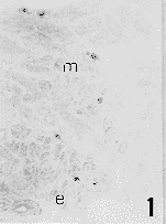
, http://www.100md.com
图版说明
图1 孕0d小鼠子宫壁内CD57+细胞的分布,阳性细胞稀少,位于肌层。M.子宫肌层 E.子宫内膜(下同)×l50
图2 孕2d小鼠子宫壁内CD57+细胞的分布,阳性细胞散在于子宫肌层 ×150
图3 孕4d小鼠子宫壁内CD57+细胞的分布,D.蜕膜区(下同)×150
图4 孕8d小鼠于宫壁内CD57+细胞的分布,仅在肌层见到单个阳性细胞调 ×150
图5 孕6d妊娠失败小鼠子宫壁内CD57+细胞的分布,大量阳性细胞存在于肌层和内膜中 ×150
Explanation of figures
, 百拇医药
Fig. 1 CD57+ cells in the mouse uterus on day 0 of pregnancy. A few positive cells were localized in the myometrium. M, myometriurn. E,endometrium (same in the following figures). ×150
Fig. 2 CD57+ cells in the mouse uterus on day 2 of pregnancy. Positive cclls were interspersed in M. ×150
Fig. 3 CD57+ cells in the mouse uterus on day 4 of gestation. Positive cells were localized in the undecidualized E and M. D,decidua. ×150
, 百拇医药
Fig. 4 CD57+ cells in the uterus on day & of normal pregnancy. Only one positive cell was seen in the Figure. ×150
Fig. 5 CD57+ cells in the uterus on day 6 of a pregnancy failure animal. Positivc cells werc numerous and evenly distributed. ×150
* 国家自然科学基金(No.39770543)、北京市自然科学基金(6982014)及河北省自然科学基金资助课题(No.399131)
△ Department of Animal Anatomy and Embryology, College of Veterinary Medicine, China Agricultural University, Beijing 100094,China
, http://www.100md.com
作者简介:钟秀会(现工作单位:河北农业大学动物科技学院,保定 071001)
参考文献
1 Head J R. Uterine natural killer cells during pregnancy in rodents. Nat Immun, 1996-97,15(1):7
2 Baines M G, Duclos A J, Antecka E, et al. Decidual infiltration and activation of macrophages leads to early embryo loss. Am J Reprod Immunol, 1997,37(6):471
3 De Fougerolles A R, Baines M G. Modulation of the natural killer cell activity in pregnant mice alters the spontaneous abortion rate. J Reprod Immunol, 1987,11(1):147
, 百拇医药
4 Chaouat G, Menu E, Clark DA, et al. Control of fetal survival in CBA×DBA/2 mice by lymphokine therapy. J Reprod Fertil, 1990,89(5):447
5 Gendron R L, Baines MG. Infiltrating decidual natural killer cells are associated with spontaneous abortion in mice. Cell Immunol, 1988,113(2):261
收稿1998-08 修回1999-03, 百拇医药
单位:中国农业大学动物医学院解剖学组织胚胎学教研室,北京 100094
关键词:NK细胞;妊娠早期;妊娠失败;小鼠
解剖学报990424 【摘要】 目的 探索CD57+自然杀伤细胞(NK细胞,natural killer cells)在小鼠妊娠早期子宫壁内的意义。方法 用小鼠抗人NK细胞CD57单克隆抗体,对未经产小鼠妊娠早期子宫壁内NK细胞进行免疫组织化学标记。结果 孕0d,小鼠子宫壁内见少量CD57+细胞,孕2d时NK细胞数量剧增,孕4d时稍下降。孕6d、孕8d时数量更少(低于0d值)。孕6d和8d均出现妊娠失败个体,其子宫壁内NK细胞很多,显著高于正常妊娠6d和8d小鼠的数量(P<0.01)。从分布区域看,胚胎着床(4.5d)前,NK细胞多位于子宫肌层,胚胎着床后仅存在于着床点以外的子宫内膜和肌层。在妊娠失败个体,NK细胞广泛存在于子宫内膜和肌层中。 结论 小鼠交配后子宫NK细胞增多与子宫炎性反应有关。着床期以后子宫NK细胞增多与妊娠失败有一定的相关性。
, 百拇医药
DISTRIBUTION OF CD57+ NATURAL KILLER CELLS IN
MOUSE UTERUS DURING EARLY PREGNANCY
Zhong Xiuhui, Zhou Zhanxiang△,Sun Binggui, Deng Zepei, Liu Jiwu
(Department of Animal Anatomy and Embryology, College of Veterinary Medicine, China Agricultural University, Beijing)
【Abstract】 Objective To realize the significance of CD57+ natural killer cells in mouse uterus during early pregnancy. Methods Using a mouse-anti-human CD57 monoclonal antibody to localize NK cells in uterus of virgin mice. Results There are a few CD57+ cells in the uterus on day 0 of pregnancy. The NK cells are elevated sharply in number on day 2 and become less on day 4. On days 6 and 8 of normal pregnancies, the cell numbers are much decreased(lower than the value of day 0). At this time, pregnancy failure was found in some individuals, in which the CD57+ cells occur in great numbers in the uteri. In reference to distribution, the NK cells are usually found in the myometrium prior to implantation (day 4.5). NK cells only occur in the undecidualized endometrium or myometrium post implantation in normal pregnancies. In pregnancy failure,however, the cells are evenly distributed in whole uterus.Conclusion The NK cells in mouse uterus increase after mating, implying the participation of NK cells in the inflammatory response of the uterus to mating and the elevation of NK cells in the uterus after the time of implantation is associated with pregnancy failure.
, 百拇医药
【Key words】 Natural killer cells; Early pregnancy; Pregnancy failure; Mouse
哺乳动物妊娠过程中,胚胎着床成功与否受母体免疫环境的直接影响,子宫NK细胞是其中重要的免疫组分。近年来,以鼠类作为动物模型,对NK细胞在妊娠子宫中的分布、增殖以及功能做了较详尽的研究[1],但对NK细胞在胚胎着床过程中的意义仍不清楚。
研究小鼠NK细胞国外常用的抗体有NK1.1、Asialo-GM1,以及近年来研制的泛NK细胞单克隆抗体DX5等。对于小鼠抗人CD57+单克隆抗体,虽有技术资料(美国NeoMarkers公司)介绍能够识别小鼠、大鼠、仓鼠和鸡的NK细胞,但迄今国内外未见试验研究报道。本试验用CD57单抗研究了NK细胞在妊娠早期小鼠子宫壁内的分布,以探索CD57+细胞在妊娠早期小鼠子宫壁内存在的意义。
, http://www.100md.com
材料和方法
1.动物及取材
8周龄未经产昆明种小鼠。常规饲养,自由采食饮水。每日光照12h。经适应1周后阴道涂片法作发情检查,发情者与公鼠(1∶1)同笼过夜,次日早晨检查有阴栓者为孕0d。取材共分7组,每组小鼠5只。孕0、2、4d各1组。孕6d、孕8d各取2组。6A、8A为妊娠正常组。子宫角外观有串珠状隆起,胚胎个数清晰可见。后经切片组织学检查胚胎发育正常。6B、8B为胚胎丢失组,子宫角比未孕子宫角粗大些,未见明显胚胎隆起。切片中见到蜕膜化子宫内膜未完全复旧。
颈椎脱臼法处死孕鼠,剖腹取出子宫,波恩液固定48h,梯度酒精脱水,Paraplast(美国Sigma公司)包埋。滑动切片机纵切(厚6μm)。
2.免疫组织化学染色
切片脱蜡入水后,经H2O2封闭,抗原热修复,进行免疫组织化学染色。主要程序如下:10%犊牛血清封闭,滴加CD57单克隆抗体(北京邦定生物医学公司),4℃过夜,生物素化羊抗鼠IgM(美国Vector公司)37℃孵育30min,HRP标记链霉亲和素(life technologies,Inc.)37℃孵育30min,每步骤之间用0.01mol/L PBS(pH7.4)冲洗3次。0.03%DAB显色5~10min,常规脱水透明,封片。
, 百拇医药
3.细胞计数
40倍物镜下观察,每样品取20个视野,统计阳性细胞总数,计算组内个体间的平均值。t检验法计算组间差异显著性。
结果和讨论
孕0d,子宫壁内见少量CD57+细胞,散在于肌层(图1)。孕2d时,CD57+细胞增多,从0d时的16.3个增加到62.3个(P<0.01),仍分布于子宫肌层(图2)。有些个体可见CD57+细胞成簇存在于子宫内膜和内环肌之间。孕2d时子宫NK细胞增多,可能与交配引起的子宫炎性反应有关,说明NK细胞在胚胎着床前可能起着抗感染作用。
孕4d时,子宫内膜蜕膜化,进入胚胎着床期(4.5d完成着床)。此时子宫壁内CD57+细胞比孕2d时减少。但仍高于0d(P<0.01)。细胞存在于着床点以外未蜕膜化的子宫内膜和肌层中(图3)。这显示,胚胎植入不依赖NK细胞的存在。
, 百拇医药
孕6d和孕8d剖杀的小鼠出现两种情况,一种是妊娠正常者,子宫角外观有串珠状隆起,内含发育正常的胚胎。此时子宫壁内CD57+细胞很少,位于胚胎着床点以外的肌层(图4)。说明CD57+细胞不是胚胎存活和生长所需要的细胞成分[2]。
孕6d和孕8d时的第2种情况是出现妊娠失败个体。子宫角内没有胚胎,看不到串珠状隆起,仅比未孕子宫角粗大些。子宫内CD57+细胞很多,遍布于子宫内膜和肌层中(图5)。表明子宫壁内NK细胞增多与妊娠失败有密切关系。虽然本试验结果不能直接证明,子宫壁内NK细胞增多参与或直接引起妊娠失败的发生,但从前人研究结果来看,抑制NK细胞活性能显著降低孕鼠胚胎吸收率[3],激活NK细胞活性则增加胚胎吸收率[4],说明NK细胞增多是诱发胚胎吸收的原因。加拿大学者Gendron和Baines[5]也发现,子宫壁内NK细胞增多是小鼠发生早期自发性流产的诱因。由此我们有理由推论,本试验观察到的小鼠孕6d、孕8d时的妊娠失败,与子宫壁内CD57+NK的细胞增多有关。
, http://www.100md.com
CD57+细胞在不同妊娠日龄小鼠子宫壁内的数量分布及组间差异性比较结果见附表。
附表 小鼠妊娠早期子宫内CD57+细胞的分布
Table CD57+ cells in mouse uterus in early pregnancy 妊娠日龄
day of pregnancy
0d
2d
4d
6d
8d
, http://www.100md.com 正常妊娠
normal pregnancy
16.3±
2.4
62.3±
7.6a,b
33±
5.2a,b
9.7±
1.3
7.3±
1.3
, http://www.100md.com
妊娠失败
pregnancy failure
65.7±
7.2c
64.6±
4.8c
注:表中数字为×40物镜下CD57+细胞总数(每样品均计20个视野)
a:与0d比,P<0.01,b:与6d,8d(正常妊娠组)比,P<0.01,c:与相应日龄正常妊娠组比较,P<0.01)
Notes:The figures in Table are the total numbers of CD57+ cells of 20 high power fields.a,P<0.01, compared with day 0 of pregnancy.b,P<0.01 compared with those of day 6 and day 8 of normal pregnancy.c,P<0.01 in comparison with the normal pregnancies on day 6 or 8,respectively.


, http://www.100md.com
图版说明
图1 孕0d小鼠子宫壁内CD57+细胞的分布,阳性细胞稀少,位于肌层。M.子宫肌层 E.子宫内膜(下同)×l50
图2 孕2d小鼠子宫壁内CD57+细胞的分布,阳性细胞散在于子宫肌层 ×150
图3 孕4d小鼠子宫壁内CD57+细胞的分布,D.蜕膜区(下同)×150
图4 孕8d小鼠于宫壁内CD57+细胞的分布,仅在肌层见到单个阳性细胞调 ×150
图5 孕6d妊娠失败小鼠子宫壁内CD57+细胞的分布,大量阳性细胞存在于肌层和内膜中 ×150
Explanation of figures
, 百拇医药
Fig. 1 CD57+ cells in the mouse uterus on day 0 of pregnancy. A few positive cells were localized in the myometrium. M, myometriurn. E,endometrium (same in the following figures). ×150
Fig. 2 CD57+ cells in the mouse uterus on day 2 of pregnancy. Positive cclls were interspersed in M. ×150
Fig. 3 CD57+ cells in the mouse uterus on day 4 of gestation. Positive cells were localized in the undecidualized E and M. D,decidua. ×150
, 百拇医药
Fig. 4 CD57+ cells in the uterus on day & of normal pregnancy. Only one positive cell was seen in the Figure. ×150
Fig. 5 CD57+ cells in the uterus on day 6 of a pregnancy failure animal. Positivc cells werc numerous and evenly distributed. ×150
* 国家自然科学基金(No.39770543)、北京市自然科学基金(6982014)及河北省自然科学基金资助课题(No.399131)
△ Department of Animal Anatomy and Embryology, College of Veterinary Medicine, China Agricultural University, Beijing 100094,China
, http://www.100md.com
作者简介:钟秀会(现工作单位:河北农业大学动物科技学院,保定 071001)
参考文献
1 Head J R. Uterine natural killer cells during pregnancy in rodents. Nat Immun, 1996-97,15(1):7
2 Baines M G, Duclos A J, Antecka E, et al. Decidual infiltration and activation of macrophages leads to early embryo loss. Am J Reprod Immunol, 1997,37(6):471
3 De Fougerolles A R, Baines M G. Modulation of the natural killer cell activity in pregnant mice alters the spontaneous abortion rate. J Reprod Immunol, 1987,11(1):147
, 百拇医药
4 Chaouat G, Menu E, Clark DA, et al. Control of fetal survival in CBA×DBA/2 mice by lymphokine therapy. J Reprod Fertil, 1990,89(5):447
5 Gendron R L, Baines MG. Infiltrating decidual natural killer cells are associated with spontaneous abortion in mice. Cell Immunol, 1988,113(2):261
收稿1998-08 修回1999-03, 百拇医药