侵袭性血管粘液瘤——附4例临床病理分析
作者:廖晓耘 李 荣 回允中
单位:北京医科大学人民医院病理科,北京 100044
关键词:侵袭性血管粘液瘤☆;外阴肿瘤;病理学;间叶瘤;病理学;粘液瘤;病理学
北京医科大学学报990428 摘 要 目的:分析侵袭性血管粘液瘤的临床和病理学特点。方法:对1988年3月~1998年6月我院收治的4例侵袭性血管粘液瘤患者的临床资料进行回顾性分析,并对其病理切片行免疫组化染色。结果:4例均为女性,年龄39~50岁;3例发生在外阴,1例在盆腔底部。肿瘤最大径4~15 cm。免疫组化染色肿瘤细胞阳性反应分别为vimentin (4/4)、desmin (4/4)、 actin (1/4)、SMA (1/4)、CD34(3/4), S-100蛋白为阴性;雌激素受体、孕激素受体全部呈强阳性表达。随访4例均无复发和转移。结论:侵袭性血管粘液瘤是一种少见的特征性的间叶性肿瘤,主要累及成年女性的会阴和盆腔,肿瘤呈浸润性生长,但不发生转移。肿瘤具有鲜明的病理组织学特点,免疫组化显示肿瘤细胞可能为纤维母细胞/肌纤维母细胞来源,在其生长过程中激素可能起一定的作用。
, 百拇医药
中国图书资料分类法分类号 R732.12
Aggressive angiomyxoma : a clinicopathologic study of 4 cases
LIAO Xiao-Yun, LI Rong, HUI Yun-Zhong
(Department of Pathology, People's Hospital, Beijing Medical University, Beijing 100044)
ABSTRACT Objective: To investigate the clinical and pathologic features of aggressive angiomyxoma(AA). Methods: 4 cases histologically confirmed as AA and treated in our hospital from Mar.1988 to Jun.1998 were retrospectively studied. In the meanwhile, the immunohistochemistry was utilized to further characterize the neoplasms. Results: All the 4 patients were females, aged between39 and 50 years; the soft tissues of vulva in 3 patients and pelvis floor in 1 patient were involved. The immunoreactivities of the neoplastic cells were vimentin (4/4), desmin (4/4), actin (1/4), SMA (1/4), CD34 (3/4), and S-100 protein negative; All of the tumors were strongly positive for estrogen and progesterone receptor. Follow-up studies ranged from 2~124 months, and no patient developed recurrence and metastases. Conclusions: Aggressive angiomyxoma is a rare, distinctive mensenchymal neoplasm characterized by locally infiltrative growth and a lack of metastases. The major sites of involvement are vulva and pelvis floor. The immunohistochemistry of the neoplastic cells exhibits some of fibroblastic and myofibroblastic features, and they appear to be hormonally influenced.
, 百拇医药
MeSH Aggressive angiomyxoma☆ Vulva neoplasms/pathol Mesenchymona/pathol Myxoma/pathol
侵袭性血管粘液瘤(aggressive angiomyxoma, AA)是一种少见的、主要发生在成年女性外阴和盆腔的间叶性肿瘤,其特点为局部浸润生长,术后易于复发,但从不发生转移。本病国内鲜有报告。本文对我院1988年3月~1998年6月间收治的4例侵袭性血管粘液瘤患者资料进行临床和病理分析,并对全部病例的病理切片应用SP法进行免疫组化染色,选用的抗体包括vimentin, desmin, actin, SMA,CD34, S-100蛋白, estrogen receptor(ER)和progesterone receptor(PR),以作进一步定性分析。
1 临床资料
4例均为女性,年龄39~50岁,平均年龄45.5岁;均为首次发病。3例发生在外阴,并向深部生长,其中1例肿瘤上达阴道穹窿顶端,下至坐骨结节;1例在盆腔底部。病程半年至11年不等。前3例表现为无痛性肿物,后1例为治疗子宫腺鳞癌手术中偶然发现。3例外阴肿物行肿瘤切除术,术后未辅以其它治疗;合并子宫腺鳞癌者除行子宫加双附件切除,并清扫盆腔淋巴结外,一并切除盆底肿物,术后镭照射2次。4例全部随访,时间为2~124月,均健在,未发现局部复发和远处转移。
, http://www.100md.com
2 病理检查
2.1 大体形态
肿瘤最大径4~15 cm。2例为不规则实性条索状肿物;1例呈分叶状,无明显包膜;1例则为扁平舌状,有蒂,有部分包膜。肿物质韧,半透明状,切面灰白、黄白或粉红色,部分呈褐色。3例呈水肿粘液变性,其中1例伴有囊性改变。
2.2 镜下所见
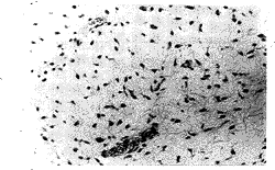
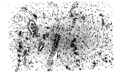
在疏松、水肿或粘液样的胶原纤维性间质中,可见两种基本成份:疏松的间叶性肿瘤细胞和丰富的血管(图1)。肿瘤细胞为梭形或星形,胞浆少,淡染;细胞核为圆形或椭圆形,均匀细小,染色质致密,核仁不明显(图2)。灶状细胞成份增多,细胞较肥胖,但未见核分裂像和细胞非典型性。血管成份是由不同大小的动脉、静脉和毛细血管构成,散在或随机分布在整个粘液性基质中。部分区域可见血管呈簇状分布以及中度肥大增厚的动脉,但未见吻合和分枝的血管。血管扩张充血,3例可见血管周围灶状出血。2例可见肿瘤浸润成熟脂肪组织,1例动脉周围可见小的分散的平滑肌束。
, http://www.100md.com
图1 于粘液样基质中疏松分布的梭形细胞和不同大小的血管
是侵袭性血管粘液瘤的诊断特点。苏木素-伊红染色 ×100
Figure 1 Spindled tumor cells and blood vessels of variable caliber
loosely distributed in a myxoid matrix are diagnostic
of aggressive angiomyxoma. Hematoxylin and eosin ×100
2.3 免疫组化结果
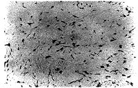
4例肿瘤细胞vimentin、desmin(图3)、ER、PR均呈强阳性,3例CD34阳性, 1例SMA、actin阳性,S-100蛋白未见阳性表达(表1)。
, 百拇医药
3 讨论
侵袭性血管粘液瘤于1983年由Steeper等[1]首次报道,英语文献报告不足100例[2];男∶女比例约为1∶6[3]。女性一般见于绝经期前的中年妇女,发病部位有外阴、阴道、盆腔底部、直肠周围、腹股沟、臀部、后腹膜;男性发生于阴囊、精索、腹股沟、肛周和盆腔;由于肿瘤较大,常可侵及几个部位[1~4]。本组女性4例,年龄39~50岁,平均年龄45.5岁,均发生在外阴、阴道穹窿及盆腔底部。
图2 肿瘤细胞呈梭形或星形,细胞学形态良好。
细胞核小,卵圆形,均匀一致,染色质致密,核仁不明显。
未见核多形性和分裂像。苏木素-伊红染色 ×200
, http://www.100md.com
Figure 2 The tumor cells are spindled or stellate-shaped and cytologically
bland. They have small, oval, uniform nuclei, with dense chromatin
and indistinct nucleoli. Nuclear pleomorphism and mitotic figures
were absent. Hematoxylin and eosin ×200
临床上常表现为无痛性肿物,在腹股沟和盆底还可表现为疝[4]。病程半年到11年不等,术前常诊断为巴氏腺囊肿、阴道囊肿、腹股沟疝等。
病理诊断上,侵袭性血管粘液瘤需要与下述良性或恶性的富于粘液或水肿性基质的间叶性肿瘤鉴别。(1)血管肌纤维母细胞瘤是一种少见的良性外阴软组织肿瘤。其特点为边界清楚,术后无复发;镜下间质水肿性,细胞丰富和细胞稀少的区域交替出现,肿瘤细胞梭形至卵圆形,聚集在薄壁毛细血管周围;缺乏AA中的厚壁血管。此肿瘤于近年才陆续报告[5]。(2)浅表性血管粘液瘤是一种不常见的皮肤和皮下肿瘤,发生于躯干、四肢、头颈部,呈单个或多结节状生长,边界清楚,易复发,无AA的厚壁血管。(3)肌间粘液瘤最常发生于成年人的股部、肩部和上肢的大肌肉间,细胞成份更少,缺乏血管,含有丰富的酸性粘液。(4)粘液性神经纤维瘤的肿瘤细胞核为细长弯曲波纹状,神经性标记物S-100蛋白阳性,无AA中的明显的血管成份。(5)恶性肿瘤肿瘤细胞有异型性、核分裂像及坏死等形态学特点。此外,由于各种间叶性肿瘤的组织来源不同,仔细寻找也可以找到其特异性分化的证据。例如,粘液性脂肪肉瘤存在脂肪母细胞和显著编织状结构的分枝毛细血管,缺乏AA中的不同大小的血管。阴道葡萄状肉瘤多见于幼女,偶尔可见横纹肌母细胞及肌浆内横纹。
, http://www.100md.com
免疫组化,肿瘤细胞vimentin和desmin均强阳性,支持肿瘤细胞可能起源于间质细胞,并表现出纤维母细胞和肌纤维母细胞的一些特点。
表1 免疫组化结果
Table 1 Immunohistochemical results Case
Vimentin
Desmin
Actin
SMA
CD34
S-100
ER
, http://www.100md.com
PR
1
+++
+++
-
-
+
-
+++
+++
2
+++
++
-
, 百拇医药
-
+
-
+++
+++
3
+++
+++
+++
+++
+
-
+++
+++
, http://www.100md.com
4
+++
+++
-
-
-
-
+++
+++
The immunohistochemical results were evaluated by the percentage of positive stromal cells expressing antigens. -, <10%; +, 10%-30%; ++, 31%-60%; +++,>60%.
, 百拇医药
图3 肿瘤细胞免疫组化染色desmin强阳性。免疫组化SP法 ×200
Figure 3 Strong desmin immunoreactivity in stromal cells. SP ×200
近来有学者注意到侵袭性血管粘液瘤与雌激素和孕激素之间的关系,但观察结果有所不同。Sutton等[7]用被覆葡聚糖的碳分析法观察到肿瘤细胞存在等量的ER和PR;Fetsch等[4]报告的ER和PR阳性率分别为93%和90%; Htwe等[8]报告一例妊娠妇女外阴AA生长与妊娠期明显相关,免疫组化染色显示PR强阳性,而ER阴性;Havel等[9]报告ER免疫染色强阳性,而PR阴性;Westcott等[2]用免疫过氧化物酶染色发现肿瘤细胞的ER和PR均强阳性表达。本组的4例女性患者ER和PR全部强阳性,加之此肿瘤较特异的生长部位和明显的女性发病倾向,提示肿瘤进展中激素因素的作用。
, 百拇医药
治疗均采取外科切除。对手术不能彻底清除肿瘤的某些选择性病例术后可以辅以抗激素治疗[2,4,7];有学者报告术前用血管造影栓塞处理,引起肿瘤缺血,以提高病变的可视性,术前外部光照射和术中电子光束放射治疗[10];这些方法都可能起到减少局部复发的机会。
文献报告复发率高达36%~72%,首次复发可见于长达14年之后[4]。本组术后虽未见复发,但仍需进行长期随访。迄今,尚无淋巴结和远处转移的报告[1~4,6~10]。
☆ 自由词。
参考文献
1 Steeper TA, Rosai J. Aggressive angiomyxoma of the female pelvis and perineum: report of nine cases of a distinctive type of gynecologic soft tissue neoplasm. Am J Surg Pathol, 1983, 7: 463-475
, http://www.100md.com
2 Westcott CJ, Gardner R, Marks GJ. Aggressive angiomyxoma presenting as a pelvic floor hernia. Surgery, 1997, 122: 969-972
3 Iezzoni JC, Fechner RE, Wong LS, et al. Aggressive angiomyxoma in males: a report of four cases. Am J Clin Pathol, 1995, 104: 391-396
4 Fetsch JF, Laskin WB, Lefkowitz M, et al. Aggressive angiomyxoma: a clinicopathologic study of 29 female patients. Cancer, 1996, 78: 79-90
, 百拇医药
5 Fletcher CDM, Tsang WYW, Fisher C, et al. Angiomyofibroblastoma of the vulva: A benign neoplasm distinct from aggressive angiomyxoma. Am J Surg Pathol, 1992, 16: 373-382
6 Begin LR, Clement PB, Kirk ME, et al. Aggressive angiomyxoma of pelvic soft parts: a clinicopathologic study of nine cases. Hum Pathol, 1985, 16: 621-628
7 Sutton GP, Rogers RE, Roth LM, et al. Aggressive angioyxoma first diagnosed as levator hernia. Am J Obstet Gynecol, 1989, 161: 73-75
, 百拇医药
8 Htwe M, Deppisch LM, Saint-Julien JS. Hormone-dependent, aggressive angiomyxoma of the vulva. Obstet Gynecol, 1995, 86(4pt2): 697-699
9 Havel G, Burian P, Kohrtz M, et al. Aggressive angiomyxoma of the vulva: an unusual, deceptive and recurrence prone tumor with evidence of estrogen receptor expression. APMIS, 1994, 102: 236-240
10 Nyam DCNK, Pemberton JH. Large aggressive angiomyxoma of the perineum and pelvis: an alternative approach, report of a case. Dis Colon Rectum, 1998, 41: 514-516
(1998-09-16收稿), 百拇医药
单位:北京医科大学人民医院病理科,北京 100044
关键词:侵袭性血管粘液瘤☆;外阴肿瘤;病理学;间叶瘤;病理学;粘液瘤;病理学
北京医科大学学报990428 摘 要 目的:分析侵袭性血管粘液瘤的临床和病理学特点。方法:对1988年3月~1998年6月我院收治的4例侵袭性血管粘液瘤患者的临床资料进行回顾性分析,并对其病理切片行免疫组化染色。结果:4例均为女性,年龄39~50岁;3例发生在外阴,1例在盆腔底部。肿瘤最大径4~15 cm。免疫组化染色肿瘤细胞阳性反应分别为vimentin (4/4)、desmin (4/4)、 actin (1/4)、SMA (1/4)、CD34(3/4), S-100蛋白为阴性;雌激素受体、孕激素受体全部呈强阳性表达。随访4例均无复发和转移。结论:侵袭性血管粘液瘤是一种少见的特征性的间叶性肿瘤,主要累及成年女性的会阴和盆腔,肿瘤呈浸润性生长,但不发生转移。肿瘤具有鲜明的病理组织学特点,免疫组化显示肿瘤细胞可能为纤维母细胞/肌纤维母细胞来源,在其生长过程中激素可能起一定的作用。
, 百拇医药
中国图书资料分类法分类号 R732.12
Aggressive angiomyxoma : a clinicopathologic study of 4 cases
LIAO Xiao-Yun, LI Rong, HUI Yun-Zhong
(Department of Pathology, People's Hospital, Beijing Medical University, Beijing 100044)
ABSTRACT Objective: To investigate the clinical and pathologic features of aggressive angiomyxoma(AA). Methods: 4 cases histologically confirmed as AA and treated in our hospital from Mar.1988 to Jun.1998 were retrospectively studied. In the meanwhile, the immunohistochemistry was utilized to further characterize the neoplasms. Results: All the 4 patients were females, aged between39 and 50 years; the soft tissues of vulva in 3 patients and pelvis floor in 1 patient were involved. The immunoreactivities of the neoplastic cells were vimentin (4/4), desmin (4/4), actin (1/4), SMA (1/4), CD34 (3/4), and S-100 protein negative; All of the tumors were strongly positive for estrogen and progesterone receptor. Follow-up studies ranged from 2~124 months, and no patient developed recurrence and metastases. Conclusions: Aggressive angiomyxoma is a rare, distinctive mensenchymal neoplasm characterized by locally infiltrative growth and a lack of metastases. The major sites of involvement are vulva and pelvis floor. The immunohistochemistry of the neoplastic cells exhibits some of fibroblastic and myofibroblastic features, and they appear to be hormonally influenced.
, 百拇医药
MeSH Aggressive angiomyxoma☆ Vulva neoplasms/pathol Mesenchymona/pathol Myxoma/pathol
侵袭性血管粘液瘤(aggressive angiomyxoma, AA)是一种少见的、主要发生在成年女性外阴和盆腔的间叶性肿瘤,其特点为局部浸润生长,术后易于复发,但从不发生转移。本病国内鲜有报告。本文对我院1988年3月~1998年6月间收治的4例侵袭性血管粘液瘤患者资料进行临床和病理分析,并对全部病例的病理切片应用SP法进行免疫组化染色,选用的抗体包括vimentin, desmin, actin, SMA,CD34, S-100蛋白, estrogen receptor(ER)和progesterone receptor(PR),以作进一步定性分析。
1 临床资料
4例均为女性,年龄39~50岁,平均年龄45.5岁;均为首次发病。3例发生在外阴,并向深部生长,其中1例肿瘤上达阴道穹窿顶端,下至坐骨结节;1例在盆腔底部。病程半年至11年不等。前3例表现为无痛性肿物,后1例为治疗子宫腺鳞癌手术中偶然发现。3例外阴肿物行肿瘤切除术,术后未辅以其它治疗;合并子宫腺鳞癌者除行子宫加双附件切除,并清扫盆腔淋巴结外,一并切除盆底肿物,术后镭照射2次。4例全部随访,时间为2~124月,均健在,未发现局部复发和远处转移。
, http://www.100md.com
2 病理检查
2.1 大体形态
肿瘤最大径4~15 cm。2例为不规则实性条索状肿物;1例呈分叶状,无明显包膜;1例则为扁平舌状,有蒂,有部分包膜。肿物质韧,半透明状,切面灰白、黄白或粉红色,部分呈褐色。3例呈水肿粘液变性,其中1例伴有囊性改变。
2.2 镜下所见
在疏松、水肿或粘液样的胶原纤维性间质中,可见两种基本成份:疏松的间叶性肿瘤细胞和丰富的血管(图1)。肿瘤细胞为梭形或星形,胞浆少,淡染;细胞核为圆形或椭圆形,均匀细小,染色质致密,核仁不明显(图2)。灶状细胞成份增多,细胞较肥胖,但未见核分裂像和细胞非典型性。血管成份是由不同大小的动脉、静脉和毛细血管构成,散在或随机分布在整个粘液性基质中。部分区域可见血管呈簇状分布以及中度肥大增厚的动脉,但未见吻合和分枝的血管。血管扩张充血,3例可见血管周围灶状出血。2例可见肿瘤浸润成熟脂肪组织,1例动脉周围可见小的分散的平滑肌束。

, http://www.100md.com
图1 于粘液样基质中疏松分布的梭形细胞和不同大小的血管
是侵袭性血管粘液瘤的诊断特点。苏木素-伊红染色 ×100
Figure 1 Spindled tumor cells and blood vessels of variable caliber
loosely distributed in a myxoid matrix are diagnostic
of aggressive angiomyxoma. Hematoxylin and eosin ×100
2.3 免疫组化结果
4例肿瘤细胞vimentin、desmin(图3)、ER、PR均呈强阳性,3例CD34阳性, 1例SMA、actin阳性,S-100蛋白未见阳性表达(表1)。
, 百拇医药
3 讨论
侵袭性血管粘液瘤于1983年由Steeper等[1]首次报道,英语文献报告不足100例[2];男∶女比例约为1∶6[3]。女性一般见于绝经期前的中年妇女,发病部位有外阴、阴道、盆腔底部、直肠周围、腹股沟、臀部、后腹膜;男性发生于阴囊、精索、腹股沟、肛周和盆腔;由于肿瘤较大,常可侵及几个部位[1~4]。本组女性4例,年龄39~50岁,平均年龄45.5岁,均发生在外阴、阴道穹窿及盆腔底部。

图2 肿瘤细胞呈梭形或星形,细胞学形态良好。
细胞核小,卵圆形,均匀一致,染色质致密,核仁不明显。
未见核多形性和分裂像。苏木素-伊红染色 ×200
, http://www.100md.com
Figure 2 The tumor cells are spindled or stellate-shaped and cytologically
bland. They have small, oval, uniform nuclei, with dense chromatin
and indistinct nucleoli. Nuclear pleomorphism and mitotic figures
were absent. Hematoxylin and eosin ×200
临床上常表现为无痛性肿物,在腹股沟和盆底还可表现为疝[4]。病程半年到11年不等,术前常诊断为巴氏腺囊肿、阴道囊肿、腹股沟疝等。
病理诊断上,侵袭性血管粘液瘤需要与下述良性或恶性的富于粘液或水肿性基质的间叶性肿瘤鉴别。(1)血管肌纤维母细胞瘤是一种少见的良性外阴软组织肿瘤。其特点为边界清楚,术后无复发;镜下间质水肿性,细胞丰富和细胞稀少的区域交替出现,肿瘤细胞梭形至卵圆形,聚集在薄壁毛细血管周围;缺乏AA中的厚壁血管。此肿瘤于近年才陆续报告[5]。(2)浅表性血管粘液瘤是一种不常见的皮肤和皮下肿瘤,发生于躯干、四肢、头颈部,呈单个或多结节状生长,边界清楚,易复发,无AA的厚壁血管。(3)肌间粘液瘤最常发生于成年人的股部、肩部和上肢的大肌肉间,细胞成份更少,缺乏血管,含有丰富的酸性粘液。(4)粘液性神经纤维瘤的肿瘤细胞核为细长弯曲波纹状,神经性标记物S-100蛋白阳性,无AA中的明显的血管成份。(5)恶性肿瘤肿瘤细胞有异型性、核分裂像及坏死等形态学特点。此外,由于各种间叶性肿瘤的组织来源不同,仔细寻找也可以找到其特异性分化的证据。例如,粘液性脂肪肉瘤存在脂肪母细胞和显著编织状结构的分枝毛细血管,缺乏AA中的不同大小的血管。阴道葡萄状肉瘤多见于幼女,偶尔可见横纹肌母细胞及肌浆内横纹。
, http://www.100md.com
免疫组化,肿瘤细胞vimentin和desmin均强阳性,支持肿瘤细胞可能起源于间质细胞,并表现出纤维母细胞和肌纤维母细胞的一些特点。
表1 免疫组化结果
Table 1 Immunohistochemical results Case
Vimentin
Desmin
Actin
SMA
CD34
S-100
ER
, http://www.100md.com
PR
1
+++
+++
-
-
+
-
+++
+++
2
+++
++
-
, 百拇医药
-
+
-
+++
+++
3
+++
+++
+++
+++
+
-
+++
+++
, http://www.100md.com
4
+++
+++
-
-
-
-
+++
+++
The immunohistochemical results were evaluated by the percentage of positive stromal cells expressing antigens. -, <10%; +, 10%-30%; ++, 31%-60%; +++,>60%.

, 百拇医药
图3 肿瘤细胞免疫组化染色desmin强阳性。免疫组化SP法 ×200
Figure 3 Strong desmin immunoreactivity in stromal cells. SP ×200
近来有学者注意到侵袭性血管粘液瘤与雌激素和孕激素之间的关系,但观察结果有所不同。Sutton等[7]用被覆葡聚糖的碳分析法观察到肿瘤细胞存在等量的ER和PR;Fetsch等[4]报告的ER和PR阳性率分别为93%和90%; Htwe等[8]报告一例妊娠妇女外阴AA生长与妊娠期明显相关,免疫组化染色显示PR强阳性,而ER阴性;Havel等[9]报告ER免疫染色强阳性,而PR阴性;Westcott等[2]用免疫过氧化物酶染色发现肿瘤细胞的ER和PR均强阳性表达。本组的4例女性患者ER和PR全部强阳性,加之此肿瘤较特异的生长部位和明显的女性发病倾向,提示肿瘤进展中激素因素的作用。
, 百拇医药
治疗均采取外科切除。对手术不能彻底清除肿瘤的某些选择性病例术后可以辅以抗激素治疗[2,4,7];有学者报告术前用血管造影栓塞处理,引起肿瘤缺血,以提高病变的可视性,术前外部光照射和术中电子光束放射治疗[10];这些方法都可能起到减少局部复发的机会。
文献报告复发率高达36%~72%,首次复发可见于长达14年之后[4]。本组术后虽未见复发,但仍需进行长期随访。迄今,尚无淋巴结和远处转移的报告[1~4,6~10]。
☆ 自由词。
参考文献
1 Steeper TA, Rosai J. Aggressive angiomyxoma of the female pelvis and perineum: report of nine cases of a distinctive type of gynecologic soft tissue neoplasm. Am J Surg Pathol, 1983, 7: 463-475
, http://www.100md.com
2 Westcott CJ, Gardner R, Marks GJ. Aggressive angiomyxoma presenting as a pelvic floor hernia. Surgery, 1997, 122: 969-972
3 Iezzoni JC, Fechner RE, Wong LS, et al. Aggressive angiomyxoma in males: a report of four cases. Am J Clin Pathol, 1995, 104: 391-396
4 Fetsch JF, Laskin WB, Lefkowitz M, et al. Aggressive angiomyxoma: a clinicopathologic study of 29 female patients. Cancer, 1996, 78: 79-90
, 百拇医药
5 Fletcher CDM, Tsang WYW, Fisher C, et al. Angiomyofibroblastoma of the vulva: A benign neoplasm distinct from aggressive angiomyxoma. Am J Surg Pathol, 1992, 16: 373-382
6 Begin LR, Clement PB, Kirk ME, et al. Aggressive angiomyxoma of pelvic soft parts: a clinicopathologic study of nine cases. Hum Pathol, 1985, 16: 621-628
7 Sutton GP, Rogers RE, Roth LM, et al. Aggressive angioyxoma first diagnosed as levator hernia. Am J Obstet Gynecol, 1989, 161: 73-75
, 百拇医药
8 Htwe M, Deppisch LM, Saint-Julien JS. Hormone-dependent, aggressive angiomyxoma of the vulva. Obstet Gynecol, 1995, 86(4pt2): 697-699
9 Havel G, Burian P, Kohrtz M, et al. Aggressive angiomyxoma of the vulva: an unusual, deceptive and recurrence prone tumor with evidence of estrogen receptor expression. APMIS, 1994, 102: 236-240
10 Nyam DCNK, Pemberton JH. Large aggressive angiomyxoma of the perineum and pelvis: an alternative approach, report of a case. Dis Colon Rectum, 1998, 41: 514-516
(1998-09-16收稿), 百拇医药