大脑皮质机械性损伤诱导Nestin表达和神经前体细胞增殖
作者:阮奕文 王传恩 童健尔 姚志彬
单位:中山医科大学人体解剖学教研室; 广州, 510089
关键词:大脑皮质;损伤;神经前体细胞;巢蛋白
中山医科大学学报/990303
摘要 目的:探讨皮质损伤后Nestin的表达和神经前体细胞的增殖情况。方法:在立体定位仪上横切成年大鼠的大脑皮质和胼胝体,于术后14 d和30 d分别杀死动物。用Nestin、GFAP免疫组化和Nissl染色方法,观察损伤后神经前体细胞和反应性星形胶质细胞的反应模式。结果:损伤后,伤口周围的皮质、扣带和胼胝体出现大量的Nestin阳性细胞,伤侧侧脑室室管膜下区(SVZ)的神经前体细胞也分裂增殖。Nestin在皮质的表达呈现一个以切口为中心的梯度反应模式并持续到术后30 d。在分布上,神经前体细胞与反应性星形胶质细胞有部分重叠。结论:机械性大脑皮质损伤可诱导切口周围和侧脑室SVZ神经前体细胞增殖,后者可能对脑损伤后的修复起重要作用。
, http://www.100md.com
中图号 R 322.8
Proliferation of Neural Progenitor Induced Following
Mechanial Injury of Rat Cortex
Ruan Yiwen Wang Chuan′en Tong Jian′er Yao Zhibin
(Department of Anatomy, Sun Yat-sen University of Medical Sciences, Guangzhou, 510089)
Abstract Objective:To explore the Nestin expression and the proliferation of neural progenitor after mechanical injury of rat cortex. Methods:The cortex and corpus callosum of adult SD rats were cut transversely in a stereotaxic apparatus and the animals were killed in the 14 day and 30 day following injury. The reaction pattern of neural progenitor and reactive astrocytes after brain damage was observed by Nestin and glial fibroblast acid protein(GFAP) immunostaining and Nissl staining.Results:There are a lot of Nestin positive cells appear around the wound in the cortex, cingulum and corpus callosum. The proliferation of the neural progenitor also can be found in the subventricular zone(SVZ) at the damaged side. Nestin expression in the cortex shows a gradient pattern around the cut and sustains to the 30 day. Some of neural progenitors overlap with reactive astrocytes at the site of injury. Conclusion:The mechanical damage of the cortex can induce the proliferation of neural progenitor around the wound and SVZ.
, http://www.100md.com
Subject headings cerebral cortex/injuries; neural progenitor; nestin
神经前体细胞亦称神经干细胞,是神经系统的祖先细胞。胚胎早期,脑内存在大量的神经前体细胞[1]。经过迁移定位后,它们最终分化成各种类型的神经元和胶质细胞。至胚胎晚期,神经前体细胞逐渐减少;出生后,大部分脑区已停止了细胞的分裂,神经前体细胞基本消失。但最近的研究发现,成年脑的某些区域如前脑的室管膜下区(SVZ)存在着较多的神经前体细胞,这些细胞分裂后沿着特定的路径向嗅球迁移,最后分化成颗粒细胞和小球周细胞[2,3]。海马齿状回在成年期也有神经前体细胞,它们不断分裂,以补充死亡的颗粒细胞[4]。研究表明海马齿状回、脊髓和视神经损伤均可诱导神经前体细胞的分裂[5~7]。这些结果提示,神经前体细胞在脑损伤后的修复中可能发挥重要的作用。为进一步探讨大脑皮质机械性损伤后诱导的神经前体细胞分裂情况,本研究通过手术切断皮质和胼胝体,观察损伤后切口周围及侧脑室SVZ神经前体细胞的分裂及其与反应性星形胶质细胞的关系。由于巢蛋白(Nestin)存在于分裂的神经前体细胞中[8],而成熟的神经元和胶质细胞不表达,故被选为神经前体细胞的标记物。
, http://www.100md.com
1 材料和方法
成年SD大鼠(350~400 g)15只,分3组(每组5只):①正常对照组;②手术损伤1组(术后14 d);③手术损伤2组(术后30 d)。②③组大鼠麻醉后,固定在立体定位仪上。参照Jeoje Paxinos和Charles Watson的大鼠全脑立体定位图谱,选出手术的3个坐标值,它们分别为前后坐标1.8 mm;横坐标2.0 mm;纵坐标3.0 mm。先用电动小圆钻钻开颅骨右侧,再将特制的双刃刀片(宽2.0 cm)插入脑组织,上下抽动10次,以达到横断皮质和胼胝体的目的。分别在手术损伤后14 d和30 d杀死大鼠。用4%的多聚甲醛固定液经左心室灌注固定。在嗅结节、视交叉和下丘脑后部3处横断切取脑块,正常组也在相应的部位切取脑块,用半导体冷冻切片机进行冠状切片,片厚40 μm。3套相邻切片分别用于Nestin,胶质原纤维酸性蛋白(GFAP)免疫组化染色和尼氏(Nissl)染色。兔抗Nestin(美国Mckay教授赠送)滴度为1∶2 000;鼠抗GFAP(Sigma公司),滴度为1∶4 000;抗生物素氧化染色试剂盒(ExtrAvidin Peroxidase Staining Kits, Sigma公司)的二抗和三抗滴度分别为1∶200。用常规的抗生素-生物素-过氧化酶复合物(ABC)免疫组化技术,切片经一抗(过夜)、二抗和三抗(分别为2 h)漂浮孵育,最后经3,3-二氨基苯联胺(DAB)呈色。
, 百拇医药
2 结 果
2.1 正常对照组
皮质、扣带和胼胝体均未观察到Nissl表达。但前脑(纹状体水平以前)侧脑室的SVZ出现Nestin阳性细胞。它们主要位于外侧壁上部及上角外上方(此部包绕纹状体上方),外侧壁下部亦有少量的阳性细胞。上部的神经前体细胞有2~4层,排列整齐,胞体较小,多为卵圆形或梭形,其突起伸向脑室和脑实质。侧脑室上壁和内侧壁的SVZ有少量的Nestin阳性细胞,排列稀疏。
2.2 手术损伤组
2.2.1 切口周围的Nestin和GFAP反应
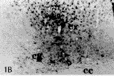
Nissl染色切片可见切口损伤的范围包括皮质、扣带和胼胝体等。术后14 d,损伤侧皮质的Nestin阳性反应最明显,涉及的范围较广。切口周围的Nestin免疫染色呈现一个梯度反应模式。离伤口越近处,反应越强烈,Nestin阳性细胞数越多;离伤口越远处,反应越弱,Nestin阳性细胞越少。皮质浅层的反应范围要比深层的大,冠状片上可观察到一个Nestin阳性的楔形反应区(图1A,见插页1)。而从前后的连续切片来看,随着逐渐远离伤口,Nestin阳性细胞的消失也逐渐是从皮质深层过渡到浅层(图1B,图1C)。在最靠近伤口的区域,Nestin阳性细胞密集,胞体肥大,多呈星形,突起粗大而长,互相交织成网,环绕着伤口的周边。离伤口稍远的部位,Nestin阳性细胞密度降低,胞体较小,突起较细而短(图2A)。非损伤侧的皮质未观察到Nestin的表达。GFAP免疫染色显示在伤口的周围出现许多胞体较大的星形阳性细胞,其出现的模式与Nestin阳性细胞的反应模式相似。但其涉及的范围、阳性细胞密度、胞体和突起的大小均小于Nestin阳性细胞(图2B)。术后30 d,皮质内切口周围仍存在大量的Nestin阳性细胞,但阳性细胞的排列较14 d的整齐(图3A)。而GFAP阳性的肥大细胞数目及其突起数目均较14 d的少(图3B)。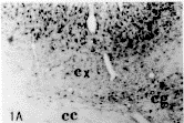

, http://www.100md.com
图1 (A,B,C)损伤后14 d大脑皮质、扣带和脐既体的Nestin阳性细胞。cx为皮质,cg为扣带,cc为饼眠体。x40
Flg.1(A,B,C)The Nestin positive cells in the cortex, clngulum and corpus calloSum in the l4 day after injury. cx (cortex), cg (cingulum), cc(corpus callosum) x40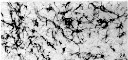
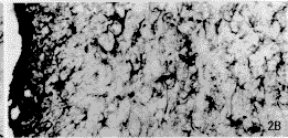
图2 损伤后14 d大脑皮质的Nestin阳性细胞汕(A)和GFAP阳性细胞(B)。左侧近切口处:右侧远离切口处。 x200
, http://www.100md.com
Flg. 2 The Nestin postive cells(A)and GFAP positive cells(B) in the cortex in the l4 day after injury。 The left side is near the cut. The right side is far from the cut. x 200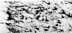
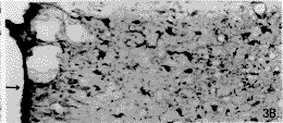
图3 损伤后30 d大脑皮质的Nestin和阳性细胞(A)和GFAP阳性细胞(B)。箭头所指为皮质的表面。x200
, http://www.100md.com Flg.3 The Nestin positive cells(A)and GFAP opsitive cells(B)in the cortex im the 30 day after injury.The arrow indicates the surfacd of the cortex.×200
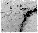
损伤侧的扣带亦可观察到与皮质出现的形态相似的Nestin阳性细胞(图1A)。非损伤侧的扣带未发现Nestin的表达。损伤侧的胼胝体内亦出现大量Nestin阳性细胞,其胞体较小,大多为梭形,这些细胞的长轴多与纤维的走向相一致(图4A)。Nestin的表达随着胼胝体的横走纤维延伸到对侧,但其阳性细胞数逐渐减少(图4B)。GFAP免疫染色显示在伤侧的扣带和胼胝体内亦出现少量胞体较大的阳性细胞。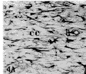
, 百拇医药
图4 损伤后14 d损伤侧(A)胼胝体的Nestin阳性细胞较多,而非伤侧(B)胼胝体的Nestin阳性细胞则较少。箭头所诣为Nestin阳性细胞。 cc为饼回氏体。 x 200
Flg.4The number of Nestin positive cells in the corpus calosum at the lesion side(A)is more than that at teh control side (B )in the 14 day after injury。the arrow indicates Nestin positive cell.cc(corpus callosum)×200
2.2.2 侧脑室SVZ内的神经前体细胞对损伤的反应
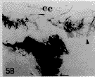
术后14 d,损伤侧的侧脑室外侧壁和上壁的SVZ中的Nestin阳性细胞增多。外侧壁的Nestin阳性细胞由正常的2~4层增加到5~7层。细胞排列不规则,胞体变大,突起增长,并有向外向上方迁移进入伤侧胼胝体的迹象(图5A)。术后30 d,SVZ中的Nestin阳性细胞虽然仍然多于非损伤侧,但少于术后14 d的Nestin阳性细胞。而非损伤侧SVZ的Nestin阳性细胞的排列与数目与正常组的大致相同(图5B)。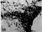
, http://www.100md.com
图5 损伤后14 d室管膜下区(A)的神经前体细胞增多,井向外向上迁移,进入伤侧胼胝体。但正常大量室营膜下区的Nestin阳性较少 (B)。细胞长箭头所指为侧脑室外侧壁:短箭头所指为上壁:CC为饼服体。x200
Fig.5The neural progenitors increase in SVZ in the 14 day after injury(A).They migrate superiorly and laterally and enter the corpus callosum in the lesion side.But the number of Nestin opsitive cells in SVZ of the normal rats is lesser than that in the damaged rats(B).T he long arrow indicates the lateral wall of the lateral ventricle and the short arrow indicates superior wall.cc(corpus callosm)×200
, 百拇医药
3 讨 论
3.1 损伤区Nestin的表达模式
本研究结果显示大脑皮质和胼胝体机械损伤后可诱导伤口周围出现大量的Nestin表达,由于Nestin可作为神经前体细胞的标志物,因而Nestin阳性细胞可被看作为神经前体细胞。Nestin在皮质的表达呈现一个以切口为中心的梯度反应模式,这可能与损伤后各部分的营养因子的浓度不同有关。体外培养实验显示:碱性成纤维细胞生长因子(bFGF)和表皮生长因子(EGF)可促进胚胎和成年的神经前体细胞分裂增殖[9]。向脑室注入bFGF同样会诱导体内SVZ和海马等区的神经前体细胞增生[10]。视网膜损伤后,可增加bFGF mRNA的表达量[11]。而产生和分泌bFGF主要来源于反应性星形胶质细胞。本实验观察到在伤口的周边出现较多肥大的GFAP阳性细胞,这些细胞属于反应性星形胶质细胞。而远离伤口处反应性星形胶质细胞减少。这样,由反应性星形胶质细胞分泌出来的GFAP,亦存在一个浓度梯度形式,由此很可能就造成了损伤后神经前体细胞以一个梯度模式出现。这种梯度模式的出现对伤口的修复是有利的。因为伤口中心是损伤最严重的部位,大量的神经前体细胞环绕着伤口周边,它们的纤维交织成网,可以减少伤口的进一步扩大,有利于伤口的修复。
, 百拇医药
3.2 Nestin表达与神经前体细胞的关系
本研究结果提示皮质损伤后出现的Nestin阳性细胞中可能有两类神经前体细胞。第1类是能分化成胶质细胞的神经前体细胞,其来源可能主要是反应性胶质细胞。它们进一步可分为反应性星形胶质细胞和反应性少突胶质细胞。前者可能主要分布在切口的周围。本实验结果显示,Nestin阳性细胞与反应性星形胶质细胞在分布上有部分重叠;由于后者具有分裂的特性,亦能表达Nestin[12],故重叠的这部分Nestin阳性细胞很可能就是反应性星形胶质细胞。反应性星形胶质细胞最终分化成星形胶质细胞。反应性少突胶质细胞可能主要分布在受损的纤维束中。Carrol等人[7]于1998年报道,在去髓鞘损伤视神经模型上可诱导视神经内静止的少突胶质前体细胞分裂。本实验在胼胝体观察到的Nestin阳性细胞,其形态与反应性星形胶质细胞有所不同,其胞体较小呈梭形和三角形,细胞的长轴与纤维走向平行。这些Nestin阳性细胞,很可能是反应性少突胶质细胞。它们最终将分化成少突胶质细胞。第2类神经前体细胞很可能来自侧脑室的SVZ的神经前体细胞。许多资料已报道成年期侧脑室的SVZ中存在大量的神经前体细胞[2,3]。Frisen等人发现切断大鼠脊髓后索后,脊髓中央管的SVZ神经前体细胞出现分裂,并向损伤区迁移[6]。本实验亦观察到损伤侧的SVZ的神经前体细胞增生并向损伤区迁移的迹象。此类神经前体细胞从侧脑室的SVZ迁移到损伤区,有可能分化为胶质细胞和神经元。因此,它们在中枢神经系统损伤后修复中的意义非常重要。Gold[5]报道了用神经兴奋性毒鹅膏覃氨酸(Ibotenic acid)和机械损伤齿状回颗粒层,可诱导齿状回的神经前体细胞增生,它们最终分化成颗粒细胞。皮质的神经元损伤溃变后是否能出现新生的神经元,需要进一步的深入研究。
, 百拇医药
参考文献
1.Dahlstrand J, Lardelli M, Lendahl U. Nestin mRNA expression correlates with the central nervous system progenitor cell state in many, but not all, regions of developing central nervous system. Brain Res Dev Brain Res, 1995,84(1):109
2.Hauke C, Ackermann I, Korr H. Cell proliferation in the subependymal layer of the adult mouse in vivo and in vitro. Cell Prolif, 1995,28:595
3.Lois C, Garcia Verdugo J M,Alvarez-Buylla A. Chain migration of neuronal precursors. Science, 1996,271:978
, 百拇医药
4.Cameron H A, Wooly C S, McEwen B S, et al. Differentiation of newly born neurons and glia in the dentate gyrus of the adult rat. Neuroscience, 1993,56(2):337
5.Gold E, Tanapat P. Lesion-induced proliferation of neuronal progenitors in the dentate gyrus of the adult rat. Neuroscience, 1997,80(2):427
6.Frisen J, Johansson C B, Torok C, et al. Rapid, widespread, and long-lasting induction of Nestin contributes to the generation of glial scar tissue after CNS injury. J Cell Biol, 1995, 131(2):453
, 百拇医药
7.Carrol W M, Jennings A R, Ironside L J. Identification of the adult resting progenitor cell by autoradiographic tracking of oligodendrocyte precursors in experimental CNS demyelination. Brain, 1998,21(pt 2):293
8.Lendahl U, Zimmerman L B, McKay R D. CNS stem cells express a new class of intermediate filament protein. Cell, 1990,60(4):585
9.Critti A, Cova L, Parati E A, et al. Basic fibroblast growth factor supports the proliferation of epidermal growth factor-generated neuronal precursor cells of the adult mouse CNS. Neurosci Lett, 1995,185(3):151
, 百拇医药
10.Craig C G, Tropepe V, Morshed C M, et al. In vivo growth factor expansion of endogenous subependymal neural precursor cell populations in the adult mouse brain. J Neurosci, 1996,16(8):2649
11.Cao W, Wen R, Li F,et al. Mechanical injury increases bFGF and CNTE mRNA expression in the mouse retina. Exp Eye Res, 1997,65(2):241
12.Lin R C S, Matesic D F, Marvin M, et al. Re-expression of the intermediate filament Nestin reactive astrocytes. Neurobiol Dis, 1995,2(2):79
1999 - 01 - 25收稿
1999 - 04 - 15修回
, http://www.100md.com
单位:中山医科大学人体解剖学教研室; 广州, 510089
关键词:大脑皮质;损伤;神经前体细胞;巢蛋白
中山医科大学学报/990303
摘要 目的:探讨皮质损伤后Nestin的表达和神经前体细胞的增殖情况。方法:在立体定位仪上横切成年大鼠的大脑皮质和胼胝体,于术后14 d和30 d分别杀死动物。用Nestin、GFAP免疫组化和Nissl染色方法,观察损伤后神经前体细胞和反应性星形胶质细胞的反应模式。结果:损伤后,伤口周围的皮质、扣带和胼胝体出现大量的Nestin阳性细胞,伤侧侧脑室室管膜下区(SVZ)的神经前体细胞也分裂增殖。Nestin在皮质的表达呈现一个以切口为中心的梯度反应模式并持续到术后30 d。在分布上,神经前体细胞与反应性星形胶质细胞有部分重叠。结论:机械性大脑皮质损伤可诱导切口周围和侧脑室SVZ神经前体细胞增殖,后者可能对脑损伤后的修复起重要作用。
, http://www.100md.com
中图号 R 322.8
Proliferation of Neural Progenitor Induced Following
Mechanial Injury of Rat Cortex
Ruan Yiwen Wang Chuan′en Tong Jian′er Yao Zhibin
(Department of Anatomy, Sun Yat-sen University of Medical Sciences, Guangzhou, 510089)
Abstract Objective:To explore the Nestin expression and the proliferation of neural progenitor after mechanical injury of rat cortex. Methods:The cortex and corpus callosum of adult SD rats were cut transversely in a stereotaxic apparatus and the animals were killed in the 14 day and 30 day following injury. The reaction pattern of neural progenitor and reactive astrocytes after brain damage was observed by Nestin and glial fibroblast acid protein(GFAP) immunostaining and Nissl staining.Results:There are a lot of Nestin positive cells appear around the wound in the cortex, cingulum and corpus callosum. The proliferation of the neural progenitor also can be found in the subventricular zone(SVZ) at the damaged side. Nestin expression in the cortex shows a gradient pattern around the cut and sustains to the 30 day. Some of neural progenitors overlap with reactive astrocytes at the site of injury. Conclusion:The mechanical damage of the cortex can induce the proliferation of neural progenitor around the wound and SVZ.
, http://www.100md.com
Subject headings cerebral cortex/injuries; neural progenitor; nestin
神经前体细胞亦称神经干细胞,是神经系统的祖先细胞。胚胎早期,脑内存在大量的神经前体细胞[1]。经过迁移定位后,它们最终分化成各种类型的神经元和胶质细胞。至胚胎晚期,神经前体细胞逐渐减少;出生后,大部分脑区已停止了细胞的分裂,神经前体细胞基本消失。但最近的研究发现,成年脑的某些区域如前脑的室管膜下区(SVZ)存在着较多的神经前体细胞,这些细胞分裂后沿着特定的路径向嗅球迁移,最后分化成颗粒细胞和小球周细胞[2,3]。海马齿状回在成年期也有神经前体细胞,它们不断分裂,以补充死亡的颗粒细胞[4]。研究表明海马齿状回、脊髓和视神经损伤均可诱导神经前体细胞的分裂[5~7]。这些结果提示,神经前体细胞在脑损伤后的修复中可能发挥重要的作用。为进一步探讨大脑皮质机械性损伤后诱导的神经前体细胞分裂情况,本研究通过手术切断皮质和胼胝体,观察损伤后切口周围及侧脑室SVZ神经前体细胞的分裂及其与反应性星形胶质细胞的关系。由于巢蛋白(Nestin)存在于分裂的神经前体细胞中[8],而成熟的神经元和胶质细胞不表达,故被选为神经前体细胞的标记物。
, http://www.100md.com
1 材料和方法
成年SD大鼠(350~400 g)15只,分3组(每组5只):①正常对照组;②手术损伤1组(术后14 d);③手术损伤2组(术后30 d)。②③组大鼠麻醉后,固定在立体定位仪上。参照Jeoje Paxinos和Charles Watson的大鼠全脑立体定位图谱,选出手术的3个坐标值,它们分别为前后坐标1.8 mm;横坐标2.0 mm;纵坐标3.0 mm。先用电动小圆钻钻开颅骨右侧,再将特制的双刃刀片(宽2.0 cm)插入脑组织,上下抽动10次,以达到横断皮质和胼胝体的目的。分别在手术损伤后14 d和30 d杀死大鼠。用4%的多聚甲醛固定液经左心室灌注固定。在嗅结节、视交叉和下丘脑后部3处横断切取脑块,正常组也在相应的部位切取脑块,用半导体冷冻切片机进行冠状切片,片厚40 μm。3套相邻切片分别用于Nestin,胶质原纤维酸性蛋白(GFAP)免疫组化染色和尼氏(Nissl)染色。兔抗Nestin(美国Mckay教授赠送)滴度为1∶2 000;鼠抗GFAP(Sigma公司),滴度为1∶4 000;抗生物素氧化染色试剂盒(ExtrAvidin Peroxidase Staining Kits, Sigma公司)的二抗和三抗滴度分别为1∶200。用常规的抗生素-生物素-过氧化酶复合物(ABC)免疫组化技术,切片经一抗(过夜)、二抗和三抗(分别为2 h)漂浮孵育,最后经3,3-二氨基苯联胺(DAB)呈色。
, 百拇医药
2 结 果
2.1 正常对照组
皮质、扣带和胼胝体均未观察到Nissl表达。但前脑(纹状体水平以前)侧脑室的SVZ出现Nestin阳性细胞。它们主要位于外侧壁上部及上角外上方(此部包绕纹状体上方),外侧壁下部亦有少量的阳性细胞。上部的神经前体细胞有2~4层,排列整齐,胞体较小,多为卵圆形或梭形,其突起伸向脑室和脑实质。侧脑室上壁和内侧壁的SVZ有少量的Nestin阳性细胞,排列稀疏。
2.2 手术损伤组
2.2.1 切口周围的Nestin和GFAP反应
Nissl染色切片可见切口损伤的范围包括皮质、扣带和胼胝体等。术后14 d,损伤侧皮质的Nestin阳性反应最明显,涉及的范围较广。切口周围的Nestin免疫染色呈现一个梯度反应模式。离伤口越近处,反应越强烈,Nestin阳性细胞数越多;离伤口越远处,反应越弱,Nestin阳性细胞越少。皮质浅层的反应范围要比深层的大,冠状片上可观察到一个Nestin阳性的楔形反应区(图1A,见插页1)。而从前后的连续切片来看,随着逐渐远离伤口,Nestin阳性细胞的消失也逐渐是从皮质深层过渡到浅层(图1B,图1C)。在最靠近伤口的区域,Nestin阳性细胞密集,胞体肥大,多呈星形,突起粗大而长,互相交织成网,环绕着伤口的周边。离伤口稍远的部位,Nestin阳性细胞密度降低,胞体较小,突起较细而短(图2A)。非损伤侧的皮质未观察到Nestin的表达。GFAP免疫染色显示在伤口的周围出现许多胞体较大的星形阳性细胞,其出现的模式与Nestin阳性细胞的反应模式相似。但其涉及的范围、阳性细胞密度、胞体和突起的大小均小于Nestin阳性细胞(图2B)。术后30 d,皮质内切口周围仍存在大量的Nestin阳性细胞,但阳性细胞的排列较14 d的整齐(图3A)。而GFAP阳性的肥大细胞数目及其突起数目均较14 d的少(图3B)。


, http://www.100md.com
图1 (A,B,C)损伤后14 d大脑皮质、扣带和脐既体的Nestin阳性细胞。cx为皮质,cg为扣带,cc为饼眠体。x40
Flg.1(A,B,C)The Nestin positive cells in the cortex, clngulum and corpus calloSum in the l4 day after injury. cx (cortex), cg (cingulum), cc(corpus callosum) x40


图2 损伤后14 d大脑皮质的Nestin阳性细胞汕(A)和GFAP阳性细胞(B)。左侧近切口处:右侧远离切口处。 x200
, http://www.100md.com
Flg. 2 The Nestin postive cells(A)and GFAP positive cells(B) in the cortex in the l4 day after injury。 The left side is near the cut. The right side is far from the cut. x 200


图3 损伤后30 d大脑皮质的Nestin和阳性细胞(A)和GFAP阳性细胞(B)。箭头所指为皮质的表面。x200
, http://www.100md.com Flg.3 The Nestin positive cells(A)and GFAP opsitive cells(B)in the cortex im the 30 day after injury.The arrow indicates the surfacd of the cortex.×200
损伤侧的扣带亦可观察到与皮质出现的形态相似的Nestin阳性细胞(图1A)。非损伤侧的扣带未发现Nestin的表达。损伤侧的胼胝体内亦出现大量Nestin阳性细胞,其胞体较小,大多为梭形,这些细胞的长轴多与纤维的走向相一致(图4A)。Nestin的表达随着胼胝体的横走纤维延伸到对侧,但其阳性细胞数逐渐减少(图4B)。GFAP免疫染色显示在伤侧的扣带和胼胝体内亦出现少量胞体较大的阳性细胞。


, 百拇医药
图4 损伤后14 d损伤侧(A)胼胝体的Nestin阳性细胞较多,而非伤侧(B)胼胝体的Nestin阳性细胞则较少。箭头所诣为Nestin阳性细胞。 cc为饼回氏体。 x 200
Flg.4The number of Nestin positive cells in the corpus calosum at the lesion side(A)is more than that at teh control side (B )in the 14 day after injury。the arrow indicates Nestin positive cell.cc(corpus callosum)×200
2.2.2 侧脑室SVZ内的神经前体细胞对损伤的反应
术后14 d,损伤侧的侧脑室外侧壁和上壁的SVZ中的Nestin阳性细胞增多。外侧壁的Nestin阳性细胞由正常的2~4层增加到5~7层。细胞排列不规则,胞体变大,突起增长,并有向外向上方迁移进入伤侧胼胝体的迹象(图5A)。术后30 d,SVZ中的Nestin阳性细胞虽然仍然多于非损伤侧,但少于术后14 d的Nestin阳性细胞。而非损伤侧SVZ的Nestin阳性细胞的排列与数目与正常组的大致相同(图5B)。


, http://www.100md.com
图5 损伤后14 d室管膜下区(A)的神经前体细胞增多,井向外向上迁移,进入伤侧胼胝体。但正常大量室营膜下区的Nestin阳性较少 (B)。细胞长箭头所指为侧脑室外侧壁:短箭头所指为上壁:CC为饼服体。x200
Fig.5The neural progenitors increase in SVZ in the 14 day after injury(A).They migrate superiorly and laterally and enter the corpus callosum in the lesion side.But the number of Nestin opsitive cells in SVZ of the normal rats is lesser than that in the damaged rats(B).T he long arrow indicates the lateral wall of the lateral ventricle and the short arrow indicates superior wall.cc(corpus callosm)×200
, 百拇医药
3 讨 论
3.1 损伤区Nestin的表达模式
本研究结果显示大脑皮质和胼胝体机械损伤后可诱导伤口周围出现大量的Nestin表达,由于Nestin可作为神经前体细胞的标志物,因而Nestin阳性细胞可被看作为神经前体细胞。Nestin在皮质的表达呈现一个以切口为中心的梯度反应模式,这可能与损伤后各部分的营养因子的浓度不同有关。体外培养实验显示:碱性成纤维细胞生长因子(bFGF)和表皮生长因子(EGF)可促进胚胎和成年的神经前体细胞分裂增殖[9]。向脑室注入bFGF同样会诱导体内SVZ和海马等区的神经前体细胞增生[10]。视网膜损伤后,可增加bFGF mRNA的表达量[11]。而产生和分泌bFGF主要来源于反应性星形胶质细胞。本实验观察到在伤口的周边出现较多肥大的GFAP阳性细胞,这些细胞属于反应性星形胶质细胞。而远离伤口处反应性星形胶质细胞减少。这样,由反应性星形胶质细胞分泌出来的GFAP,亦存在一个浓度梯度形式,由此很可能就造成了损伤后神经前体细胞以一个梯度模式出现。这种梯度模式的出现对伤口的修复是有利的。因为伤口中心是损伤最严重的部位,大量的神经前体细胞环绕着伤口周边,它们的纤维交织成网,可以减少伤口的进一步扩大,有利于伤口的修复。
, 百拇医药
3.2 Nestin表达与神经前体细胞的关系
本研究结果提示皮质损伤后出现的Nestin阳性细胞中可能有两类神经前体细胞。第1类是能分化成胶质细胞的神经前体细胞,其来源可能主要是反应性胶质细胞。它们进一步可分为反应性星形胶质细胞和反应性少突胶质细胞。前者可能主要分布在切口的周围。本实验结果显示,Nestin阳性细胞与反应性星形胶质细胞在分布上有部分重叠;由于后者具有分裂的特性,亦能表达Nestin[12],故重叠的这部分Nestin阳性细胞很可能就是反应性星形胶质细胞。反应性星形胶质细胞最终分化成星形胶质细胞。反应性少突胶质细胞可能主要分布在受损的纤维束中。Carrol等人[7]于1998年报道,在去髓鞘损伤视神经模型上可诱导视神经内静止的少突胶质前体细胞分裂。本实验在胼胝体观察到的Nestin阳性细胞,其形态与反应性星形胶质细胞有所不同,其胞体较小呈梭形和三角形,细胞的长轴与纤维走向平行。这些Nestin阳性细胞,很可能是反应性少突胶质细胞。它们最终将分化成少突胶质细胞。第2类神经前体细胞很可能来自侧脑室的SVZ的神经前体细胞。许多资料已报道成年期侧脑室的SVZ中存在大量的神经前体细胞[2,3]。Frisen等人发现切断大鼠脊髓后索后,脊髓中央管的SVZ神经前体细胞出现分裂,并向损伤区迁移[6]。本实验亦观察到损伤侧的SVZ的神经前体细胞增生并向损伤区迁移的迹象。此类神经前体细胞从侧脑室的SVZ迁移到损伤区,有可能分化为胶质细胞和神经元。因此,它们在中枢神经系统损伤后修复中的意义非常重要。Gold[5]报道了用神经兴奋性毒鹅膏覃氨酸(Ibotenic acid)和机械损伤齿状回颗粒层,可诱导齿状回的神经前体细胞增生,它们最终分化成颗粒细胞。皮质的神经元损伤溃变后是否能出现新生的神经元,需要进一步的深入研究。
, 百拇医药
参考文献
1.Dahlstrand J, Lardelli M, Lendahl U. Nestin mRNA expression correlates with the central nervous system progenitor cell state in many, but not all, regions of developing central nervous system. Brain Res Dev Brain Res, 1995,84(1):109
2.Hauke C, Ackermann I, Korr H. Cell proliferation in the subependymal layer of the adult mouse in vivo and in vitro. Cell Prolif, 1995,28:595
3.Lois C, Garcia Verdugo J M,Alvarez-Buylla A. Chain migration of neuronal precursors. Science, 1996,271:978
, 百拇医药
4.Cameron H A, Wooly C S, McEwen B S, et al. Differentiation of newly born neurons and glia in the dentate gyrus of the adult rat. Neuroscience, 1993,56(2):337
5.Gold E, Tanapat P. Lesion-induced proliferation of neuronal progenitors in the dentate gyrus of the adult rat. Neuroscience, 1997,80(2):427
6.Frisen J, Johansson C B, Torok C, et al. Rapid, widespread, and long-lasting induction of Nestin contributes to the generation of glial scar tissue after CNS injury. J Cell Biol, 1995, 131(2):453
, 百拇医药
7.Carrol W M, Jennings A R, Ironside L J. Identification of the adult resting progenitor cell by autoradiographic tracking of oligodendrocyte precursors in experimental CNS demyelination. Brain, 1998,21(pt 2):293
8.Lendahl U, Zimmerman L B, McKay R D. CNS stem cells express a new class of intermediate filament protein. Cell, 1990,60(4):585
9.Critti A, Cova L, Parati E A, et al. Basic fibroblast growth factor supports the proliferation of epidermal growth factor-generated neuronal precursor cells of the adult mouse CNS. Neurosci Lett, 1995,185(3):151
, 百拇医药
10.Craig C G, Tropepe V, Morshed C M, et al. In vivo growth factor expansion of endogenous subependymal neural precursor cell populations in the adult mouse brain. J Neurosci, 1996,16(8):2649
11.Cao W, Wen R, Li F,et al. Mechanical injury increases bFGF and CNTE mRNA expression in the mouse retina. Exp Eye Res, 1997,65(2):241
12.Lin R C S, Matesic D F, Marvin M, et al. Re-expression of the intermediate filament Nestin reactive astrocytes. Neurobiol Dis, 1995,2(2):79
1999 - 01 - 25收稿
1999 - 04 - 15修回
, http://www.100md.com