大鼠松果腺内生长抑素免疫反应神经纤维的观察
作者:徐胜春 李光武 何娟娟
单位:安徽医科大学解剖学教研室, 合肥 230032
关键词:松果体;解剖学和组织学;生长抑素;神经纤维;大鼠
安徽医科大学学报000208
摘要 目的 观察大鼠松果腺内生长抑素免疫反应(SS-IR)神经纤维的分布,为研究松果腺的神经免疫调节提供形态学依据。方法 采用ABC免疫细胞化学染色。结果 大鼠松果腺内有SS-IR神经纤维和细胞分布。腺表面及实质内均可见细长的或膨体状的阳性纤维,实质内阳性纤维分支形成网络状,以近腺茎处纤维密度相对较高,另小动脉、小静脉的周围亦有阳性纤维伴行。腺内还可见散在的SS-IR阳性细胞。结论 大鼠松果腺内含有与高等脊椎动物相同的特异性抗原,因而具有神经免疫调节的形态学基础。
中图分类号 R322.55
, 百拇医药
文献标识码 A 文章编号 1000-1492(2000)02-0106-03
Observation of somatostatin immunoreactive nerve
fibers in the pineal gland of rats
Xu Shengchun,Li Guangwu,He Juanjuan
(Dept of Anatomy,Anhui Medical University,Heifei 230032)
Abstract Objective To provide some morphological basis for neuroimmunomodulation study by observation of somatostatin immunoreactive(SS-IR) nerve fibers in the pineal gland of rats.Methods Immunohistochemical ABC method was used.Results SS-IR nerve fibers were located in both intraparenchyma and perivascular region. At the proximal end of the gland,the nerve fibers were more numerous than in the distal end.Conclusion The pineal gland of rat may contain the same specific antigen as that in the higher forms of vertebrate,showing morphological basis of neuroimmunomodulation.
, http://www.100md.com
MeSH pineal body/anat; somatostatin; nerve fibers; rats
近年来,随着神经内分泌学研究的不断深入,人们对松果腺的研究越来越多,已经确定松果腺具有多方面生理功能。国外学者〔1~8〕对啮齿类动物及牛的松果腺内生长抑素免疫反应(somatostatin immunoreaction,SS-IR)神经纤维的分布及其来源进行过研究,国内尚未见报道。本实验采用ABC免疫细胞化学方法系统地观察了大鼠松果腺内生长抑素能纤维的分布及特点,为松果腺的生理功能及药理学研究提供形态学依据。
1 材料与方法
取SD大鼠10只(♀♂不限) ,用2%戊巴比妥钠(30~40 mg/kg)腹腔麻醉,行胸腹腔联合切口,暴露心脏后,从左心室快速灌注生理盐水250 ml,再用250 ml 1.33 mol.L-1多聚甲醛灌注,先快后慢,滴完为止。打开颅腔迅速取出松果腺,将标本制成7 μm厚的石蜡切片,常规脱蜡脱水,按ABC方法进行染色,步骤如下:①0.3% H2O2 ,室温,20 min;②0.3% Tirton-100,室温,30 min;③正常马血清(1∶50)孵育,室温,30 min;④兔抗SS(1∶400),4℃,72 h;⑤生物素标记的鼠抗兔血清(1∶100,TBC稀释)室温,1 h;⑥于ABC盒取A液和B液用PBS稀释(1∶400),室温,1 h;⑦DAB溶液中显色,在显微镜下观察反应适宜时,用自来水终止反应。以上每步之间,除了第4步外,均用PBS漂洗3次,每次5 min,最后脱水、透明、明胶封片,光镜下观察。用PBS和正常兔血清取代一抗孵育做对照实验。
, http://www.100md.com
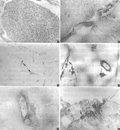
图1 大鼠松果腺的纵切面 HE×100
图2 示松果腺茎内SS-IR阳性纤维束(兼有膨体状及细长的纤维) ABC×400
图3 松果腺实质内近茎阳性纤维分支成网络状(↑) ABC×400
图4 被膜下SS-IR膨体状阳性纤维(↑)、小动脉周围的阳性纤维(A)及
阳性神经节 ( ) ABC×400
图5 腺近尾侧内小静脉周围细长的阳性纤维(V) ABC×400
图6 腺浅层内阳性神经细胞(↑) ABC×400
2 结果
, http://www.100md.com
大鼠松果腺为卵圆形实质性器官(图1)。经ABC免疫组织化学方法染色后的松果腺标本内,SS-IR神经纤维和细胞呈黑色,背景为浅黄色。我们观察到SS阳性神经纤维束主要通过松果腺茎进入腺实质内,其分支构成网络状(图2、 3)布于腺细胞周围。在腺体的浅表布有串珠状的SS阳性纤维,其末梢分支形成小的神经丛,并见由丛发出的阳性纤维进入腺体内。腺实质内小动脉及小静脉的周围亦有点线状的阳性纤维伴行(图4、5)。此外,腺内可见散在的SS阳性神经细胞(图4、6),细胞呈椭圆形、圆形或多角形,胞质内充满浓密的免疫反应物。
对照实验的标本切片均呈阴性反应。
3 讨论
3.1 Moller等〔5~8〕研究了牛松果腺内SS-IR神经纤维的分布,发现阳性纤维布于松果腺实质内及小血管的周围,阳性纤维以松果腺吻侧( 近茎处)最为密集。我们用ABC法在大鼠松果腺内所见的SS-IR阳性纤维的分布与牛松果腺相似,阳性纤维除布于血管周围外,另在腺细胞之间分支形成复杂的网络,其末梢支配松果腺细胞。从分布密度看,以吻侧较密集,其次为腺体中央部,而远侧较为稀疏。
, http://www.100md.com
3.2 近年来,随着神经内分泌学研究的不断深入,人们对松果腺内的肽能神经及其来源的研究日益增多。多数研究者〔1~3〕认为:松果腺内的肽能神经除来自颈上神经节的交感性起源外,亦有中枢起源,而且中枢起源亦有不同的组织起源。我们发现进入大鼠松果腺内SS-IR纤维大部分通过松果腺茎导入,在松果腺的表面亦见串珠状的SS-IR纤维并形成丛,其分支进入腺实质,同时在小血管的周围亦有SS-IR阳性纤维伴行。这与Peinado等〔5〕在牛等哺乳动物的松果腺内所见相似,提示大鼠松果腺内的生长抑素能纤维亦有多种来源。现已知生长抑素能抑制生长激素、促甲状腺激素等激素的释放。有学者〔6〕认为松果腺内的生长抑素可能对松果腺激素的合成与释放具有远期的影响 ,其确切的生理作用尚待进一步研究。
徐胜春,男,35岁,硕士,讲师
参考文献
1 Moller M,Korf HW.The origin of central pinealopetal nerve fibers in the Mongolian gerbil as demonstrated by the retrograde transport of horseradish peroxidase.Cell Tissue Res,1983;230(2):273~287
, http://www.100md.com
2 Mikkelsen JD,Moller M.A direct neuronal projection from the intergeniculate leaflet of the lateral geniculate nucleus to the deep pineal gland of the rat,demonstrated with phaseolus vulgaris leucoagglutinin. Brain Res,1990;520(1~2):342~346
3 Mikkelsen JD,Cozzi B, Moller M.Efferent projections from the lateral geniculate nucleus to the pineal complex of the mongolian gerbil. Cell Tissue Res,1991;264(1):95~102
4 Webb SM,Peinado MA,Puig-Domingo M et al.Rhythms in pineal immunoreactive somatostatin in the syrian hamster,mouse and gerbil. J Pineal Res,1988;5(3):273~278
, 百拇医药
5 Peinado MA,Viader M,Puig-Domingo M et al.Regional distribution of immuno reactive somatostatin in the bovine pineal gland.Neuroendocrinology,1989;50(5):550~554
6 Moller M,Mikkelsen JD,Holst JJ et al. Somatostatin and prosomatostatin immunoreactive nerve fibers in the bovinepinealgland.Neuroendocrinology,1992;56(2):278~283
7 Viader M,Mato E,Tugues D et al.In vitro and in vivo flow cytometry comparative analysis of somatostatin-positive cells in the pineal gland of the neonatal rat.Neuroendocrinology.1995;62(1):87~92
8 Mato E, Santisteban P,Chowen JA et al.Circannual somatostatin gene and somatostatin receptor gene expression in the early post-natal rat pineal gland. Neuroendocrinology.1997;66(5):368~374
1999-11-02收稿,2000-01-10修回, 百拇医药
单位:安徽医科大学解剖学教研室, 合肥 230032
关键词:松果体;解剖学和组织学;生长抑素;神经纤维;大鼠
安徽医科大学学报000208
摘要 目的 观察大鼠松果腺内生长抑素免疫反应(SS-IR)神经纤维的分布,为研究松果腺的神经免疫调节提供形态学依据。方法 采用ABC免疫细胞化学染色。结果 大鼠松果腺内有SS-IR神经纤维和细胞分布。腺表面及实质内均可见细长的或膨体状的阳性纤维,实质内阳性纤维分支形成网络状,以近腺茎处纤维密度相对较高,另小动脉、小静脉的周围亦有阳性纤维伴行。腺内还可见散在的SS-IR阳性细胞。结论 大鼠松果腺内含有与高等脊椎动物相同的特异性抗原,因而具有神经免疫调节的形态学基础。
中图分类号 R322.55
, 百拇医药
文献标识码 A 文章编号 1000-1492(2000)02-0106-03
Observation of somatostatin immunoreactive nerve
fibers in the pineal gland of rats
Xu Shengchun,Li Guangwu,He Juanjuan
(Dept of Anatomy,Anhui Medical University,Heifei 230032)
Abstract Objective To provide some morphological basis for neuroimmunomodulation study by observation of somatostatin immunoreactive(SS-IR) nerve fibers in the pineal gland of rats.Methods Immunohistochemical ABC method was used.Results SS-IR nerve fibers were located in both intraparenchyma and perivascular region. At the proximal end of the gland,the nerve fibers were more numerous than in the distal end.Conclusion The pineal gland of rat may contain the same specific antigen as that in the higher forms of vertebrate,showing morphological basis of neuroimmunomodulation.
, http://www.100md.com
MeSH pineal body/anat; somatostatin; nerve fibers; rats
近年来,随着神经内分泌学研究的不断深入,人们对松果腺的研究越来越多,已经确定松果腺具有多方面生理功能。国外学者〔1~8〕对啮齿类动物及牛的松果腺内生长抑素免疫反应(somatostatin immunoreaction,SS-IR)神经纤维的分布及其来源进行过研究,国内尚未见报道。本实验采用ABC免疫细胞化学方法系统地观察了大鼠松果腺内生长抑素能纤维的分布及特点,为松果腺的生理功能及药理学研究提供形态学依据。
1 材料与方法
取SD大鼠10只(♀♂不限) ,用2%戊巴比妥钠(30~40 mg/kg)腹腔麻醉,行胸腹腔联合切口,暴露心脏后,从左心室快速灌注生理盐水250 ml,再用250 ml 1.33 mol.L-1多聚甲醛灌注,先快后慢,滴完为止。打开颅腔迅速取出松果腺,将标本制成7 μm厚的石蜡切片,常规脱蜡脱水,按ABC方法进行染色,步骤如下:①0.3% H2O2 ,室温,20 min;②0.3% Tirton-100,室温,30 min;③正常马血清(1∶50)孵育,室温,30 min;④兔抗SS(1∶400),4℃,72 h;⑤生物素标记的鼠抗兔血清(1∶100,TBC稀释)室温,1 h;⑥于ABC盒取A液和B液用PBS稀释(1∶400),室温,1 h;⑦DAB溶液中显色,在显微镜下观察反应适宜时,用自来水终止反应。以上每步之间,除了第4步外,均用PBS漂洗3次,每次5 min,最后脱水、透明、明胶封片,光镜下观察。用PBS和正常兔血清取代一抗孵育做对照实验。

, http://www.100md.com
图1 大鼠松果腺的纵切面 HE×100
图2 示松果腺茎内SS-IR阳性纤维束(兼有膨体状及细长的纤维) ABC×400
图3 松果腺实质内近茎阳性纤维分支成网络状(↑) ABC×400
图4 被膜下SS-IR膨体状阳性纤维(↑)、小动脉周围的阳性纤维(A)及
阳性神经节 (
 ) ABC×400
图5 腺近尾侧内小静脉周围细长的阳性纤维(V) ABC×400
图6 腺浅层内阳性神经细胞(↑) ABC×400
2 结果
, http://www.100md.com
大鼠松果腺为卵圆形实质性器官(图1)。经ABC免疫组织化学方法染色后的松果腺标本内,SS-IR神经纤维和细胞呈黑色,背景为浅黄色。我们观察到SS阳性神经纤维束主要通过松果腺茎进入腺实质内,其分支构成网络状(图2、 3)布于腺细胞周围。在腺体的浅表布有串珠状的SS阳性纤维,其末梢分支形成小的神经丛,并见由丛发出的阳性纤维进入腺体内。腺实质内小动脉及小静脉的周围亦有点线状的阳性纤维伴行(图4、5)。此外,腺内可见散在的SS阳性神经细胞(图4、6),细胞呈椭圆形、圆形或多角形,胞质内充满浓密的免疫反应物。
对照实验的标本切片均呈阴性反应。
3 讨论
3.1 Moller等〔5~8〕研究了牛松果腺内SS-IR神经纤维的分布,发现阳性纤维布于松果腺实质内及小血管的周围,阳性纤维以松果腺吻侧( 近茎处)最为密集。我们用ABC法在大鼠松果腺内所见的SS-IR阳性纤维的分布与牛松果腺相似,阳性纤维除布于血管周围外,另在腺细胞之间分支形成复杂的网络,其末梢支配松果腺细胞。从分布密度看,以吻侧较密集,其次为腺体中央部,而远侧较为稀疏。
, http://www.100md.com
3.2 近年来,随着神经内分泌学研究的不断深入,人们对松果腺内的肽能神经及其来源的研究日益增多。多数研究者〔1~3〕认为:松果腺内的肽能神经除来自颈上神经节的交感性起源外,亦有中枢起源,而且中枢起源亦有不同的组织起源。我们发现进入大鼠松果腺内SS-IR纤维大部分通过松果腺茎导入,在松果腺的表面亦见串珠状的SS-IR纤维并形成丛,其分支进入腺实质,同时在小血管的周围亦有SS-IR阳性纤维伴行。这与Peinado等〔5〕在牛等哺乳动物的松果腺内所见相似,提示大鼠松果腺内的生长抑素能纤维亦有多种来源。现已知生长抑素能抑制生长激素、促甲状腺激素等激素的释放。有学者〔6〕认为松果腺内的生长抑素可能对松果腺激素的合成与释放具有远期的影响 ,其确切的生理作用尚待进一步研究。
徐胜春,男,35岁,硕士,讲师
参考文献
1 Moller M,Korf HW.The origin of central pinealopetal nerve fibers in the Mongolian gerbil as demonstrated by the retrograde transport of horseradish peroxidase.Cell Tissue Res,1983;230(2):273~287
, http://www.100md.com
2 Mikkelsen JD,Moller M.A direct neuronal projection from the intergeniculate leaflet of the lateral geniculate nucleus to the deep pineal gland of the rat,demonstrated with phaseolus vulgaris leucoagglutinin. Brain Res,1990;520(1~2):342~346
3 Mikkelsen JD,Cozzi B, Moller M.Efferent projections from the lateral geniculate nucleus to the pineal complex of the mongolian gerbil. Cell Tissue Res,1991;264(1):95~102
4 Webb SM,Peinado MA,Puig-Domingo M et al.Rhythms in pineal immunoreactive somatostatin in the syrian hamster,mouse and gerbil. J Pineal Res,1988;5(3):273~278
, 百拇医药
5 Peinado MA,Viader M,Puig-Domingo M et al.Regional distribution of immuno reactive somatostatin in the bovine pineal gland.Neuroendocrinology,1989;50(5):550~554
6 Moller M,Mikkelsen JD,Holst JJ et al. Somatostatin and prosomatostatin immunoreactive nerve fibers in the bovinepinealgland.Neuroendocrinology,1992;56(2):278~283
7 Viader M,Mato E,Tugues D et al.In vitro and in vivo flow cytometry comparative analysis of somatostatin-positive cells in the pineal gland of the neonatal rat.Neuroendocrinology.1995;62(1):87~92
8 Mato E, Santisteban P,Chowen JA et al.Circannual somatostatin gene and somatostatin receptor gene expression in the early post-natal rat pineal gland. Neuroendocrinology.1997;66(5):368~374
1999-11-02收稿,2000-01-10修回, 百拇医药