淋巴结针吸细胞病理学对淋巴结核早期诊断的研究应用
 |
王 颖 王永才 赵成艳 李海东
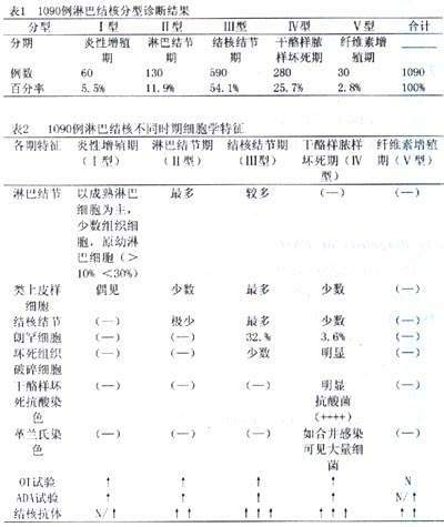
【摘要】目的 探讨淋巴结针吸细胞病理学对淋巴结核早期诊断的研究,提高淋巴结核诊断准确率。方法根据显微镜下淋巴结核不同时期的针吸细胞病理学特点及特征性结构改变,结合抗酸染色和结核抗体检测做出分型诊断。结果在1090例结核性淋巴腺炎中,男360例,占33.0%,女730例,占67.0%,年龄1~78岁,平均28.3岁。结核初期―炎性增殖期60例占5.5%;结核早期―淋巴结节期130例占11.9%;结核中期―结核性结节期有590例,占54.1%;结核晚期―干酪样脓样坏死期有280例占25.7%;结核恢复期―纤维素增殖期30例占2.8%。炎性增殖反应期非特异性形态学变化,需要结合结核抗体阳性和腺苷脱氨酶明显增高有助于诊断,结核结节期主要可有较多类上皮样细胞及郎罕氏细胞;而干酪样脓样坏死主要见大量坏死组织及碎屑、少数残碎不全类上皮样细胞,此期主要能查到抗酸菌为特点; 纤维增殖期,抽出物难取,仅见少数纤维组织、纤维细胞和粘液间质为其特征,提示结核恢复、疤痕形成所致。结论应用淋巴结针吸细胞病理学分型诊断,而且为淋巴结核早期诊断、早期发现、早期治疗提供重要方法。
, 百拇医药
关键词:淋巴结 针吸 结核 早期诊断
文章编号:1008-6919(2006)07-0068-03
中图分类号:R322.2+5
文献标识码:A
The research of the diagnosis of tuberculotu lymphadenitis typing basedon needle aspirate cytology of lymphnode
[ Abstract ] Objective To evaluate the research of needle aspirate cytology of lymphnode in the diagnosis ofthe typingoftuberculotu lymphadenitis and the function of the typing to improve the accurate rate in diagnosy .Method tuberculotu lymphadenitis type were diagnosed according to the FNA pathological characteristics and specific construction modification under the microscope,with the aid of acid-fast stain and tuberculotu antibody’s detection .Result The 1090 cases in cluded 360 males making up 33.0% ,and 730females, making up 67.0% , with age arranged from 1to 78 years old and an average age of 28.3.The results showd that 60 cases (5.5% ) were of the inflammation reaction hyperplastic stage (1) ;130 cases (11.9% ) .Lamphaticnodular stage (2) ; 590 cases (54.1% ) tuberculous nodular stage (3) ; 280 cases (25.7% ).Caseous necrosis stage (4) ; and 30 cases (2.8% ) fibrinous hyperplastic stage (5).The inflammation reaction hyperplactic stage and lymphatic nodular stage are of nonspecific morphological changes and should be diagnosed with the help of positive tuberculin test and obvious increase of Adenosine Deaninase (ADA ) ; tuberculous nodular stage has a great number of epithelioid cells, and Langhans cells, caseous necrosis stage is charteriged by a great deal of necrotic tissue and debris, a small number of fragmented epithelioid cells, and maily by antiacid bacteria fibrinous hyperplastic stage is featured by a few fibrous tissue, cells and mucous oweing to hardness to get puncture, which neans tubercalous restoration, and scar formation. ConclusionThe application offine needle aspirate cytoiology sapplies an important nethod of early diagnosis findings and trealment.
, 百拇医药
Keyword: Lymphnode Needle Tuberculosis Typing
结核病是多发病、常见病,最近几年结核的发病逐渐上升,有卷土重来之势。淋巴腺结核在淋巴结肿大疾病中, 仅次于慢性淋巴腺炎, 名列第二位, 在青少年淋巴结肿大疾病中占首位,。可单独存在或合并肺结核,肠结核,本组总结我院两年多来1090例淋巴腺结核针吸细胞学特点及分型诊断研究, 报告如下:
1.材料与方法
1.1本组收集我院门诊、住院以及外院会诊结核性淋巴腺炎1090例, 男360例占33.0% , 女730例占67.0% , 年龄1~78岁, 平均28.3岁。诊断及分型标准, 根据细胞学特征及抗酸染色结果确定。其中细胞阳性率950例占87.2% , 抗酸染色阳性率150例占13.8%,结核抗体阳性符合率65.4%。
1.2常规消毒,选用一次性10毫升塑料注射器,挑选有意义肿大淋巴结,左手固定,右手持针进行多方位穿刺抽吸取材,涂片、干后,进行瑞氏-姬姆萨双重混合染色。对干酪样坏死标本,分别做抗酸染色及革兰氏染色, 对非典型标本涂片同时做结核抗体和结核菌素试验(OT)及腺苷酸脱氨酶(ADA) 测定。显微镜下检查,根据淋巴结核不同时期的细胞学特点及特征性结构,结合抗酸染色和结核抗体的结果做出分型诊断[1、2、3、5]。
2.结果
淋巴结核分型诊断结果及各型的细胞学特征见表1,表2。
[ 下 页 ], http://www.100md.com(王 颖 王永才 等)