乳腺分叶状肿瘤的X线诊断评价(2)
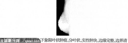 |
总之,在X线摄影片上表现圆形或椭圆形,分叶状,实性肿块,边缘完整,部分有透明晕环绕,无钙化,无肿大淋巴结显示,与乳腺纤维腺瘤有时难以鉴别,结合年龄较大,乳腺突然发现较大肿瘤以及短期内迅速增大的肿瘤,要考虑乳腺分叶状肿瘤的可能。
参考文献
[1] Liberman L, Bonaccio E, Hamele-Bena D, et al.Benign and M alignant Phyllodes Tumors: Mammographic and Sonographic Findings.Radiology , 1996, 198 (1): 121-124.
[2] 巴明臣, 崔书中, 唐云强,等. 乳腺叶状囊肉瘤24例分析.临床外科杂志,2007,15(3):167-168.
[3] 王洪江,王忠裕.国外医学(外科分册),2005,32(4):291-293.
[4] Reinfuss M, Mitus J, Duda K, et al. The treatment and prognosis of patients with phyllodes tumor of the breast. Cancer, 1996,77: 910-916.
[5] Kessinger A, Foley JF, Lemon HM, et al. Metastatic cystosarcoma phyllodes: a case report and review of the literature. J Surg Oncol, 1972, 4: 131-147.
[6]Mallebre B, Ebert A, Perez CA,et al. Cystosarcoma phyllodes of the breast. Geburshife Grauenheikd, 1996, 56(1):35-40.
[7] Page JE, Williams JE. The radiological features of phyllodes tumor of the breast with clinic-pathological correlation. Clini Radio, 1991, 157(4): 8-12.
[8] De Roos WK, Kaye P, Dent DM. Factors leading to local recurrence or death after surgical resection of phyllodes tumors of the breast. Br J Surg , 1999,86(3):396-401., http://www.100md.com(赵 虹 高洁冰 姚晋林 张翠运)